Fig. 4.
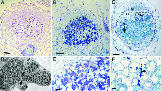
Histology of nodules with reduced DMI2 expression levels and wild-type nodules. (A) Longitudinal sections (5 μm) of 2-week-old nodules formed on (partial) DMI2 knockdown (RNAi) roots. Most cells in the central zone are filled with infection threads. (B) Longitudinal section (1 μm) of a 35S::DMI2-transformed TR25 nodule, showing wide infection threads occupying most cells of the central tissue. (C) Longitudinal section (1 μm) of a control nodule (transformed with an empty binary vector). A typical zonation of indeterminate nodules can be seen. M, meristem; zII, infection zone; zIII, fixation zone. The arrow indicates extension of the fixation zone in the direction of the nodule base. (D) Transmission electron microscopy of a 35S::DMI2-transformed TR25 nodule section, showing an infection thread inside a cell without releasing bacteria. The infection thread is surrounded by a fibrillar wall (arrowhead) and a membrane. (E) Closeup of boxed area in B, showing infection threads in the distal part of the 35S::DMI2-transformed TR25 nodule occupying major parts of the cells but with no release of bacteria. (F) Closeup of boxed area in C, showing part of the infection zone of a control nodule. Bacteria are released from infection threads, which are stained dark blue. it, infection thread. (Scale bars: A–C, 100 μm; D, 1 μm; and E and F, 20 μm.)

