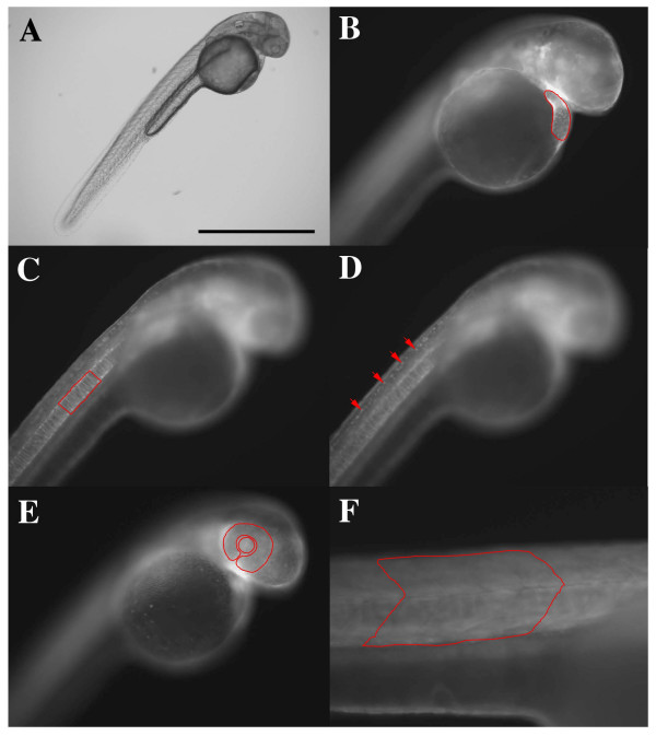
Figure 3.
Images of the same transgenic zebrafish embryo (strain: Tg(actin:egfp)zp1) 48 hours post fertilization. A. Bright field image of the embryo. B-D. Fluorescence images of the same embryo at different focal planes. B. Image at the focal plane for heart measurement. The heart is outlined in red. C. Image at the focal plane for notochord measurement. A length of notochord adjacent to 4 somites is outlined in red. D. Image at the focal plane for rohon beard neuron measurement. 4 rohon beard neurons are indicated by red arrows. E. Image at the focal plane for eye and lens measurement. The eye and lens are outlined, separately, in red. F. Image at the focal plane for somite measurement. 4 somites are outlined in red. Scale bar = 1 mm (A), 0.5 mm (B – E), 0.05 mm (F).

