
Figure 6.
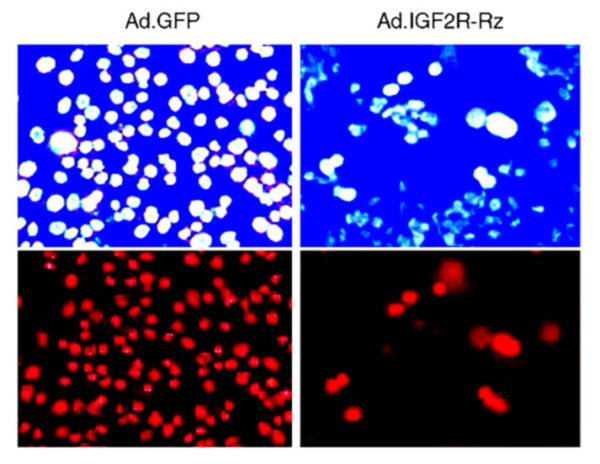
Effects of the IGF2R ribozyme on the apoptosis induced by TNF in MCF-7 cells. Cells were infected with Ad.GFP (control, left panels) or Ad.GFP/Rz-IGF2R (right panels). 72 hrs post infection, cells were treated with TNF for one day and then cell death was examined using a fluorescence microscope. Upper panels: Cells were stained with Hoechst dye for nuclei and observed under 480 nm blue-fluorescent light. The bright blue spots are the nuclei of apoptotic cells. Lower panels: Cells were stained with Propidium Iodide dye for nuclei and observed under 565 nm red-fluorescent light. The red spots are the nuclei of dead cells.