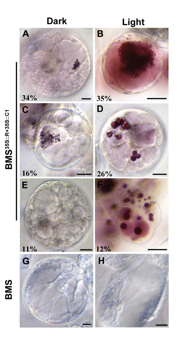
Figure 6.
Light induces alterations in the distribution of anthocyanins within vacuolar compartments. Representative DIC images of six day old (A, C, E) dark-grown and (B, D, F) light-grown BMS35S::R+35S::C1 cells. BMS cells grown in the (G) dark or (H) light show no significant alterations. Numbers correspond to the percentage values indicated in Table 2. The bar represents 20 μm.

