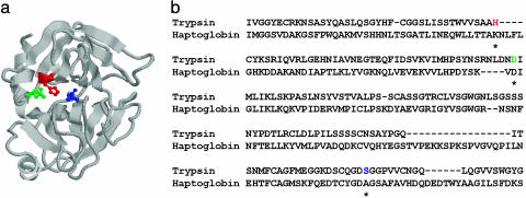
Fig. 2.
Trypsin catalytic site and trypsin sequence aligned to haptoglobin sequence. (a) Trypsin 3D structure (PDB ID code 1A0J33, chain A). Catalytic residues are shown in red (His-57), green (Asp-102), and blue (Ser-195). (b) Trypsin (PDB ID code 1A0J, chain A, residues 1–196) aligned to haptoglobin (Swiss-Prot sequence P19006, residues 85–303). Catalytic residue positions in trypsin are marked by an asterisk. 1A0J retrieved P19006 with a blast-level search; the sequences have 26% pairwise sequence identity.

