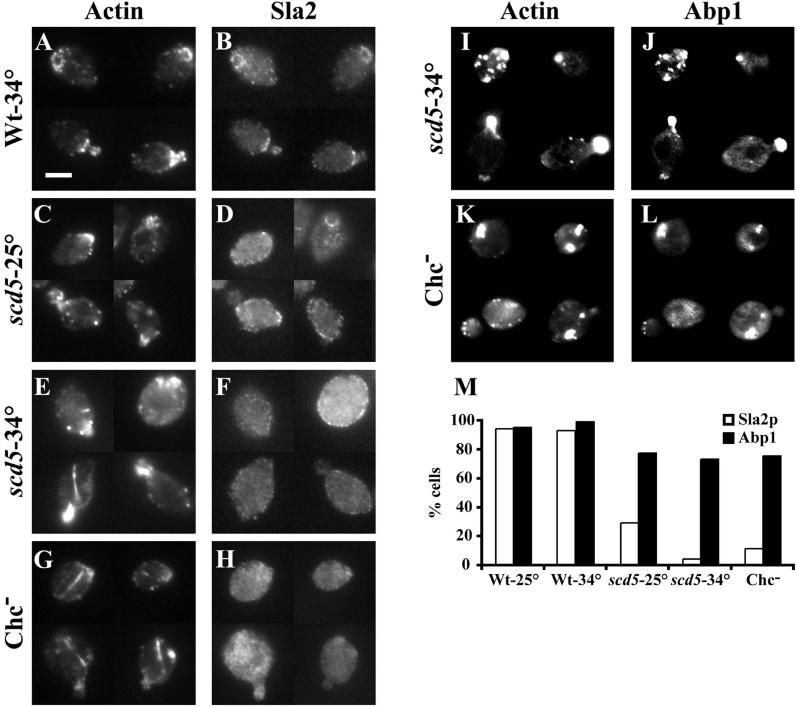
Figure 10.
Sla2p is mislocalized in scd5-Δ338 and clathrin-depleted cells. Wild-type (SL1528; A and B) and scd5-Δ338 (SL3920; C–F) strains were grown on YEPD to log phase at 25°C (C and D) or shifted to 34°C (A, B, E, F) for 3 h. The GAL1:: CHC1 strain (SL554; G and H) was grown to log phase in YEP-GAL and then shifted to YEPD for 15 h at 30°C to deplete clathrin HC. In I and J scd5-Δ338 (SL3920) + pJC4 [pGFP-ABP1] was grown in C-URA and shifted to 34°C for 3 h. In K and L the GAL1: CHC1 strain (SL554) + pJC4 [pGFP-ABP1] was grown in C-URA+GAL and then shifted to glucose medium for 15 h to deplete clathrin HC. Cells were fixed, stained with anti-actin (A, C, E, and G) and anti-Sla2p antibodies (B, D, F, and H); or in cells for GFP-Abp1p localization (J and L) actin was visualized with Alexa-594 phalloidin (I and K). (M) Quantification of the percent of cells with Sla2p or Abp1p showing colocalization with actin patches. Note that the GAL1∷CHC1 strain grown on galactose gave similar results as wild-type cells. Bar, 5 μm.

