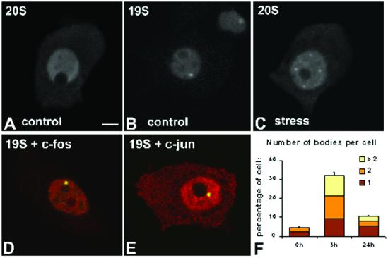
Figure 5.
Stress induces the formation of proteasome-enriched domains in rat brain neurons. Hypotalamic neurosecretory neurons were obtained from control animals (A and B) and animals sacrificed 3 h after intraperitoneal injection of an hypertonic saline solution to induce osmotic stress (C–E). Cells were immunolabeled with antibodies directed against 20S proteasome (A and C) or 19S proteasome (B). Cells were additionally double-labeled with either anti-19S proteasome (D, red staining) and anti-c-Fos antibodies (D, green staining), or anti-19S proteasome (E, red staining) and anti-c-Jun antibodies (E, green staining). (D and E) Overlay of red and green images; colocalization produces a yellowish color. Bar, 10 μm. (F) Proportion of neurons containing proteasome-enriched domains and the number of bodies per cell were estimated at 0, 3, and 24 h after saline injection. The graph depicts means ± SEs (for each time point four animals were sacrificed, and from each animal ∼150 neurons were analyzed).

