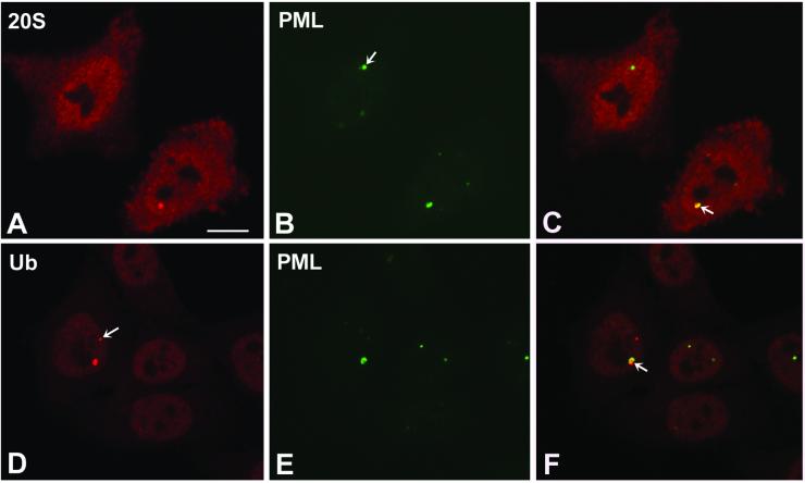
Figure 8.
PML protein is present in clastosomes. HeLa cells were double-labeled with anti-PML antibody (green staining) and either anti-20S proteasome (A and C, red staining) or anti-ubiquitin antibodies (D and F, red staining). (C and F) Overlay of red and green images; colocalization produces a yellowish color. Note that PML occupies only a fraction of large clastosomes (C and F, arrow). The arrow in B points to a PML body that does not concentrate proteasomes. In D, the arrow points to a clastosome that contains little, if any, PML. Bar, 10 μm.

