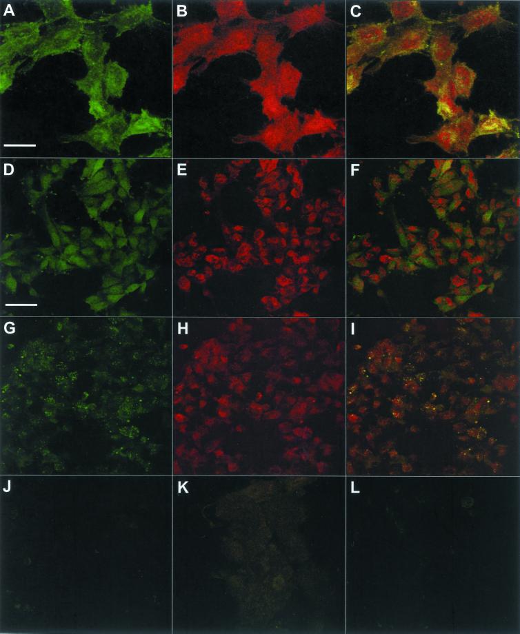
Figure 11.
Confocal microscopy of the 175-kDa HARE in SK-175HARE cells. The cellular distributions of the recombinant HARE, fl-HA, clathrin, and lysosomes were determined in SK-175HARE-34 cells as described in MATERIALS AND METHODS. (A–C) Colocalization of clathrin (A) and HARE (B) in the overlay picture (C). The different distribution patterns of HARE (D) and Lysotracker (E) in cells incubated with unlabeled HA are shown in the overlay picture (F). (I) Colocalization pattern of fl-HA (G) and Lysotracker (H). The effect of excess unlabeled HA on the uptake of fl-HA is shown in J. The background staining of SK-175HARE cells with rabbit IgG is shown in K. (L) Anti-HARE staining of SK-Hep-1 cells stably transfected with the backbone plasmid (containing no cDNA insert). The bar in A (20 μm) applies to A–C, and the bar in D (50 μm) applies to D–L.

