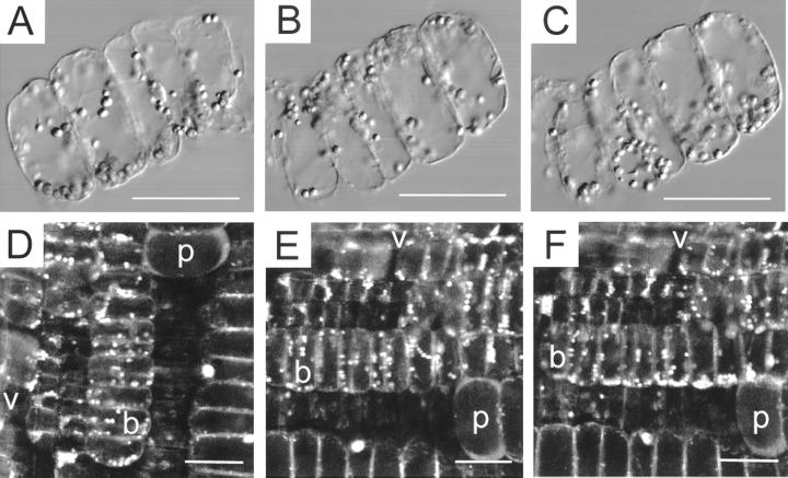
Figure 2.
Dynamics of amyloplast sedimentation. A through C, Amyloplast sedimentation in isolated files of bundle sheath cells. Amyloplasts follow the tracks of intracellular particle movement as well as the path predicted by physical parameters such as plastid density and cytosolic viscosity leading to net sedimentation of plastids to the new bottom of the cell. A, Thirty seconds before rotation of cell files through 180°. QuickTime movie located at www.plantphysiol.org. B, Thirty seconds after rotation. C, Seven minutes after rotation. D through E, Plastid sedimentation in bundle sheath cells of maize. Ratio images E2 (620–670 nm)/E1 (550–600 nm) of SNARF-1 AM-loaded longitudinal pulvinal sections, excitation 514 nm. D, Before rotation; E, 2 min after rotation by 90°. QuickTime movie located at www.plantphysiol.org. F, 12 Minutes after rotation, bar = 50 μm; v, vascular tissue; b, bundle sheath cells; p, parenchyma cells.

