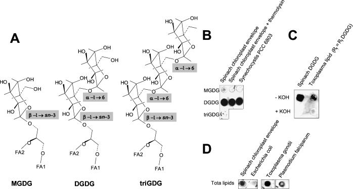
FIG. 2.
Immunodetection of DGDG in lipid extracts from T. gondii and P. falciparum. (A) Comparison of MGDG, DGDG, and triGDG polar heads. (B) The specificity of the anti-DGDG antibody for the [α-d-galactopyranosyl-(1→6)-O-β-d-galactopyranosyl[ polar head was investigated by dot blot immunostaining (1/100 dilution of anti-DGDG antibody) of 10 μg of lipids purified from spinach chloroplast envelope membranes (first two lanes) or Synechocystis PCC 6803, a cyanobacterium. The anti-DGDG antibody was detected on DGDG spots whereas it did not react with MGDG or triGDG. (C) T. gondii lipids extracted from 2 × 10⁹ free tachyzoites were separated by TLC as shown in Fig. 1. Silica at the level of the DGDG standard was scraped, and lipids were extracted (those with an Rf equal to the Rf of DGDG) and analyzed by dot blot immunostaining. The anti-DGDG antibody reacted with this spot, whereas it did not when the extracted lipids were subjected to mild alkaline hydrolysis and reextracted (+KOH). A similar immunostaining pattern was obtained with a spinach chloroplast DGDG control subjected to identical treatments. The loss of immunostaining after mild KOH hydrolysis showed that the immunostained lipids were deacylated. (D) Dot blot immunostaining with the anti-DGDG antibody of total lipid mixtures from spinach chloroplast envelope membranes (10 μg of lipids), E. coli membranes (10 μg of lipids), T. gondii (total lipid extracts from 2 × 10⁹ tachyzoites), and P. falciparum (total lipid extracts from 109 cells).

