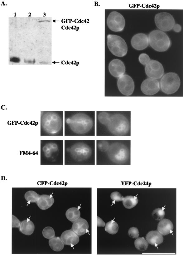
FIG. 1.
(A) GFP-Cdc42p levels were comparable to endogenous Cdc42p levels. Protein was isolated from wild-type strain C276 (lane 1), CDC42/Δcdc42 heterozygous strain DJD6-11 (lane 2), and TRY100 (lane 3), and immunoblots were probed with anti-Cdc42p antibody diluted 1:1,000. (B) GFP-Cdc42p clusters at polarized growth sites and localizes to internal membranes and the plasma membrane. TRY100 was grown in synthetic complete medium to mid-log phase and then observed for GFP-Cdc42p localization. (C) TRY100 were grown as in B, stained with FM4-64, and then viewed for colocalization of GFP-Cdc42p and vacuolar staining. (D) Colocalization of CFP-Cdc42p and YFP-Cdc24p. Plasmids p416MET(CFP-CDC42) and p415MET(YFP-CDC24) were transformed into wild-type strain TRY11-7D, and transformants were grown to mid-log phase in SC−Leu−Ura−Met medium. Arrows indicate CFP-Cdc42p localization to nuclear membranes and YFP-Cdc24p targeting to nuclei. Collages of images were assembled in Adobe Photoshop 5.0.

