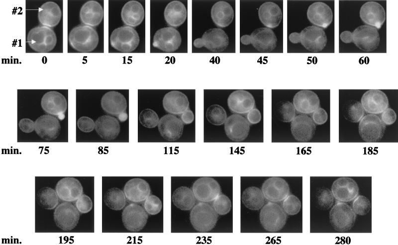
FIG. 3.
Time-lapse photomicroscopy of GFP-Cdc42p localization during the cell cycle. Wild-type haploid strain TRY11-7D was transformed with p415MET(GFP-CDC42), and transformants were grown to mid-log phase in SC−Leu−Met medium. Cells were placed onto a thin-layered agar slab made with SC−Leu−Met medium and viewed by fluorescent microscopy. GFP-Cdc42p localization was captured at ≈5- to 15-min intervals. The cells designated #1 and #2 are described in the text. These cells are representative of at least 18 cells documented with similar localization patterns.

