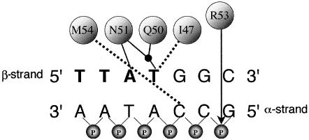
Figure 1.
Simplified representation of binding interactions between D. melanogaster Ubx protein and a double-stranded DNA target (Passner et al. 1999). At the top, I47, Q50, N51, and M54 are the four amino acids—Ile, Gln, Asn, and Met, respectively (numbered according to their position in the homeodomain)—that contact specific bases, either through hydrogen bonds (solid lines) or hydrophobic interactions (dashed lines). The black dot represents a water molecule. Designation of the α and β strands follows Billeter (1996). The 5′-TTAT-3′ motif on the β-strand of the DNA target is shown in bold. On the α-strand only, the connecting phosphate (P) groups are included to demonstrate the position (arrow) of the ionic interaction with Arg (R53). Key amino acids in human HOXD13 are identical to those in Ubx, except that M54 is replaced by V54 (Val).

