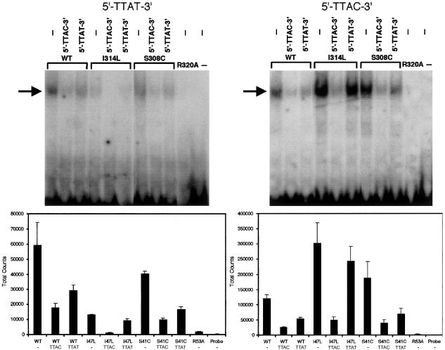
Figure 6.
In vitro binding of wild-type (WT) and mutant (I314L, S308C, and R320A) HOXD13 proteins to synthetic ds oligonucleotides. Above, representative gel shift assays employing 32P-labeled 5′-TTAT-3′ probe (left) and 5′-TTAC-3′ probe (right) in the absence (−) or presence of unlabeled competitor oligonucleotides. The arrow shows the position of the bound oligonucleotide/protein complexes. Below, quantitation (mean ± SEM) of total counts from 4–5 experiments. Note the difference in scale of absolute counts on the Y-axis.

