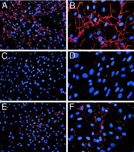
Figure 1.
Deposition of collagen VI microfibrils in the extracellular matrix of dermal fibroblasts. Fibroblasts from an unaffected control individual (A and B), patient UC-1 (C and D), and patient UC-4 (E and F) were grown in the presence of 50 μg/ml L-ascorbic acid phosphate for 4 d postconfluency and were stained with the α1(VI) collagen-specific antibody. The antibody reaction was detected with Cy3-conjugated goat anti-rabbit IgG (red), and cells were counterstained with DAPI (blue) to visualize nuclei. Original magnifications ×20 (A, C, and E) and ×40 (B, D, and F).

