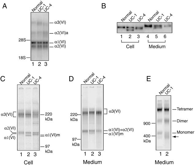
Figure 4.
Analysis of collagen VI mRNA and protein in fibroblasts from UC-1 and UC-4. A, Northern blot analysis of total RNA (5 μg) from normal fibroblasts and those from UC-1 and UC-4, with a mixture of [32P]dCTP-labeled cDNA probes for the three collagen VI mRNA. The probes hybridized with the α1(VI), α2(VI), and α3(VI) collagen mRNA of 4.2, 3.5, and 9–10 kb, respectively, plus an alternatively spliced α2(VI) collagen mRNA, α2(VI)a, of 6.0 kb. B, Immunoblot analysis of cell layer extracts (lanes 1–3) and culture media (lanes 4–6) from normal fibroblasts (lanes 1 and 4) and those from UC-1 (lanes 2 and 5) and UC-4 (lanes 3 and 6), with the antibody specific for the α1(VI) collagen chain. Lanes 1–3 contain 10 μg of total protein from the cell layers, and lanes 4–6 contain 100 μl of culture medium precipitated with 0.9 ml of 100% EtOH. C–E, Immunoprecipitation of cell layers (C) and culture media (D, E) from normal fibroblasts (lane 1) and those from UC-1 (lane 2) and UC-4 (lane 3), with the antibody specific for the α3(VI) collagen chain. Fibroblasts were labeled with [35S]cysteine overnight. Samples in B, C, and D were reduced with 25 mM DTT and separated on 3%–8% polyacrylamide gels. Samples in E were separated on a composite agarose polyacrylamide gel in the absence of 25 mM DTT. The mutant α1(VI) collagen chains are depicted as α1(VI)m. The arrow indicates a protein present in higher amounts in fibroblasts from UC-1 compared with the normal control fibroblasts.

