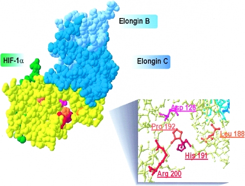
Figure 3.
VHL-polycythemia–causing and polycythemia-associated mutations. Illustration of VHL protein structure (yellow) and interactions with elongin B (pale blue), elongin C (dark blue), and HIF-1α (green) was produced by use of Swiss PDB-viewer software and was based on the PDB-1LM8 crystallized structure (Min 2002). Red, Homozygous VHL mutations found at position R200 and H191. Orange, VHL mutations found in compound heterozygosity with the 598C→T (R200W) mutation. Pink, Mutation at Y126, previously proposed as a dominant negative VHL mutation found in two siblings with apparent congenital polycythemia (Pastore et al. 2003).

