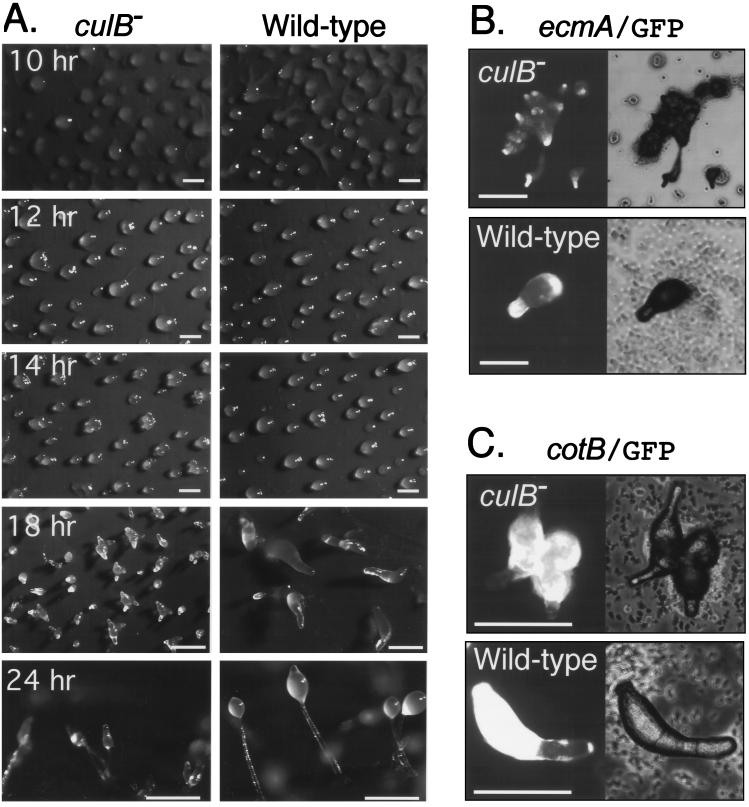
FIG. 2.
Abnormal prestalk cell differentiation in culB− cells. (A) Cells were deposited on filters and allowed to develop, and photographs were taken at the indicated times. Bars, 0.5 mm. (B and C) Cells transformed with the reporter plasmids ecmA/GFP, a prestalk marker (B), or cotB/GFP, a prespore marker (C), were photographed after 18 h of development on filters.

