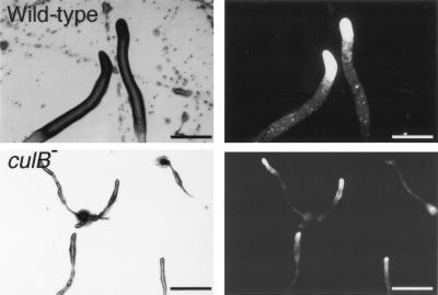
FIG. 5.
The culB− mutant is defective in slug formation and migration. Cells were deposited on nonnutrient agar plates and kept in a dark chamber with unidirectional light for 24 h. Both wild-type and culB− mutant slugs, marked with ecmA/GFP, were visualized by bright-field (left) and fluorescence (right) microscopy. Bars, 0.25 mm.

