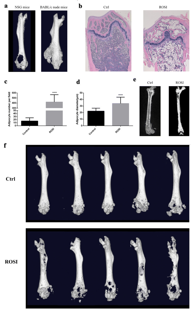
Fig. 3.
Rosiglitazone-induced marrow adipogenesis and enhanced osteolytic bone destruction. a SBC5 cells were injected into the medullary cavity of the distal femur of NSG mice and BABL/c nude mice, bone destruction was visulized using microCT. b C57BL/6J female mice were fed with diet supplemented with rosiglitazone maleate for 7 weeks. Femur was separated for HE staining. scale bars = 200 µm. Adipocyte number (c) and diameter (d) were measured using Image J. Data are represented as mean ± SD. ****p < 0.0001. e B-NSG mice were fed with diet supplemented with rosiglitazone maleate for 7 weeks. Osmium tetroxide staining was performed in the femur and microCT scanning was used to visualize and quantify BMAs. f after B-NSG mice were feed with rosiglitazone for 8 weeks, SBC5 cells were implanted by intra-femoral injection. Bone destruction was observed by microCT after 1 month

