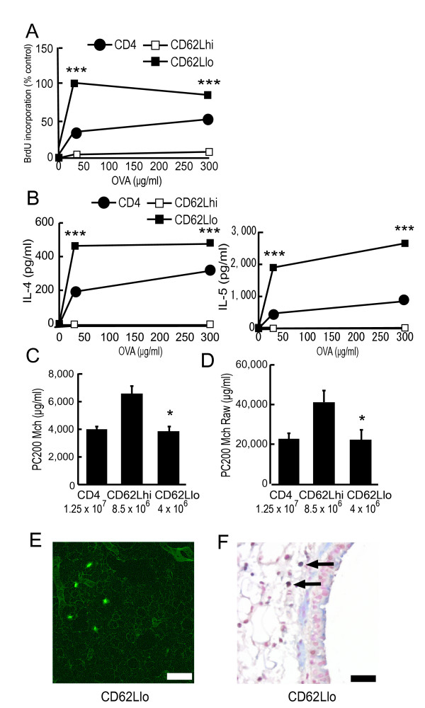
Figure 9.
CD4+CD62Llow memory/effector subset produces Th2-type cytokines and directly induces transfer-mediated AHR. (A and B)CD4+CD62Llow memory/effector subset proliferates and produces Th2-type cytokines with antigen stimulation. On day 18, CD4+ T cells (CD4), CD4+CD62Lhigh T cells (CD62Lhi), or CD4+CD62Llow T cells (CD62Llo) from OVA-sensitized mice were positively selected by magnetic cell sorting as described in Methods. Then, these cells were cultured with freshly isolated mitomycin C-treated splenocytes in the presence of OVA. After 48 hours, the proliferation was assessed by BrdU incorporation using ELISA (A). The maximum proliferation observed in response to OVA for CD4+CD62Llow T cells from OVA-sensitized mice was set as control (100%). After 72 hours, Th2-type cytokine levels in the supernatants were assayed using ELISA (B). *** p < 0.001 compared with the value of CD4+CD62Lhigh T cells. (C and D) CD4+CD62Llow memory/effector T cells induce AHR. Recipients received CD4+ (CD4; 1.25 × 10⁷), CD4+CD62Lhigh (CD62Lhi; 8.5 × 10⁶), or CD4+CD62Llow (CD62Llo; 4 × 10⁶) cells from OVA-sensitized mice. AR was measured 4 days after the transfer. (C) AR was measured by Penh methods (n = 5–6 per group). * p < 0.05 compared with PC200Mch of mice that received CD4+CD62Lhigh T cells. (D) AR was assessed by measurement of Raw (n = 6–8 per group). * p < 0.05 compared with PC200Mch Raw of mice that received CD4+CD62Lhigh T cells. (E)Fluorescence study. CD4+CD62Llow cells from OVA-sensitized mice (4 × 10⁶) were labeled with fluorescent dye, and then transferred into recipients. Five-micrometer sections of the lungs were observed by fluorescence microscopy 24 hours after the transfer (CD62Llo). Scale bar, 100 μm. (E) Double staining analysis by immunohistochemistry. T cells were detected by staining for CD3 (cytoplasm, blue). Proliferation was assessed by staining for PCNA (nucleus, brown). Lung sections from mice that received OVA-sensitized CD4+CD62Llow cells (CD62Llo; 4 × 10⁶) are shown. Proliferating T cells were clearly detected (black arrow). Scale bar, 40 μm.

