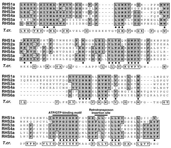
FIG. 3.
Amino acid alignment of box 1 domain from RHS1a-6a proteins and related T. cruzi protein. The aligned box 1 sequences indicated by an asterisk in the left margin of Fig. 2 are representative of each RHS subfamily. Dashes were introduced to maximize the alignment. Identical amino acids are shaded and in bold. The positions of the ATP/GTP-binding motif and the duplicated insertion site sequence associated with non-LTR retroelement (RIME and/or ingi) insertion are indicated above the alignment. The last row (T.cr.) represents the chimeric RHS-related protein identified in the T. cruzi database, as shown in Fig. 10. All boxed and bold residues of the T. cruzi sequence are identical to residues located at the same relative position in the T. brucei RHS proteins.

