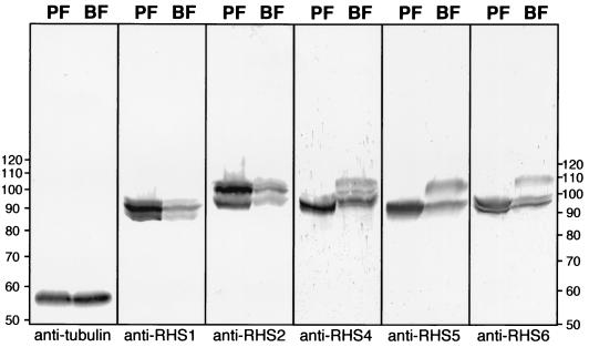
FIG. 6.
Western blot analysis of RHS proteins. Lysates (4 × 10⁷ cells) of T. brucei procyclic form EATRO1125 (PF) and bloodstream form AnTat1 (BF) were analyzed by Western blotting with the immune sera specific for tubulin, RHS1, RHS2, RHS4, RHS5, and RHS6. The positions of the molecular mass markers (in kilodaltons) are indicated on the left and right, and the names of the immune sera is given under each blot.

