Fig. 2.
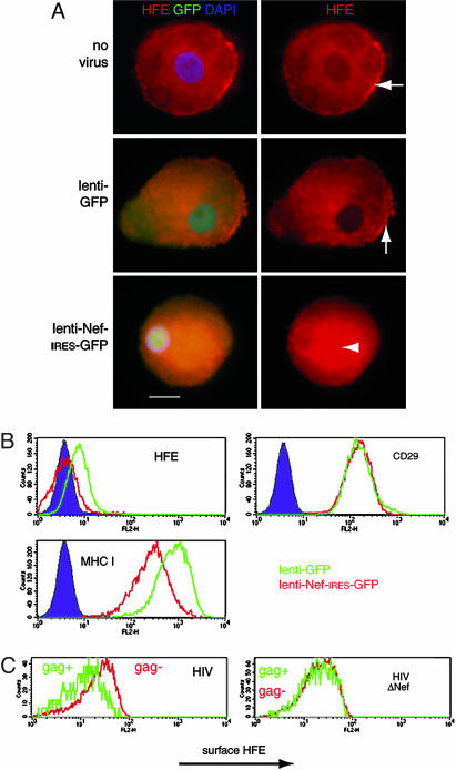
Down-regulation of endogenous surface HFE in monocytic THP-1 cells and primary macrophages by Nef and HIV. (A) Ex vivo macrophages were infected with recombinant lentivirus-based vectors encoding either GFP (lenti-GFP) or Nef-IRES-GFP (lenti-Nef-IRES-GFP) and stained for HFE (red). Images of representative cells show that surface HFE expression on membrane ruffles in uninfected cells is unaltered in cells infected with the control lenti-GFP virus (Top and Middle Right, arrows) but that HFE is redistributed to a perinuclear region in cells infected with lenti-Nef-IRES-GFP (Bottom Right, arrowhead). (Scale bar, 10 μm.) (B) The cell monocyte/macrophage cell line THP-1 normally expresses low levels of surface HFE that are unchanged by the control lenti-GFP virus, but, in cells infected with lenti-Nef-IRES-GFP, surface HFE is undetectable above the background. Nef does not alter surface CD29 (negative control) but MHC I levels are lowered (positive control). (C) Macrophages were infected for 4 days with M-tropic HIV-1, with (HIV) or without (HIVΔNef) the nef gene, stained for surface HFE (anti-α3 domain) and p18/p55 gag, and analyzed by FACS. HIV-infected gag-expressing cells (Left, green line) express less surface HFE than do uninfected cells from the same culture (Left, red line). HIVΔNef infection (Right) does not alter surface HFE. This finding was repeated on macrophages from multiple donors expressing wild-type HFE.

