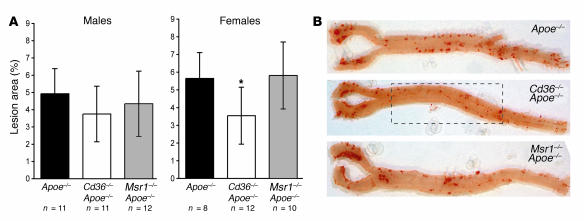
Figure 3.
Morphometric analysis of lesion area in the aortic tree. Aortae were stained en face with Sudan IV, and lesion area was measured as a percentage of total aortic area. (A) Lesion area in male and female mice. *P ≤ 0.05; significantly different from Apoe–/– control mice. (B) Representative photographs of the en face aortae from female Apoe–/–, Cd36–/–Apoe–/–, and Msr1–/–Apoe–/– mice. The boxed region indicates the area of reduced lesions observed in the Cd36–/–Apoe–/– female mice.

