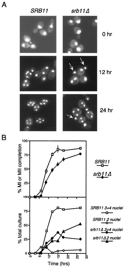
FIG. 1.
Phenotypic analysis of srb11Δ diploids during meiosis. (A) Fluorescence microscopy (magnification, ×1,000) of DAPI-stained RSY335 (SRB11) and RSY389 (srb11Δ) at 0, 12, and 24 h after transfer to sporulation medium. Arrows indicate the presence of small buds containing nucleus-sized DAPI-stained material. (B) Rate of appearance of bi- and tetranucleated cells in SRB11 and srb11Δ strains during meiosis. (Top) Percentage of cells in the culture executing at least one meiotic division, presented as a function of time following transfer to sporulation medium. MI, meiosis I; MII, meiosis II. Error bars indicate standard deviation. (Bottom) Breakdown between binucleated (2 nuclei) and tri- and tetranucleated (3+4 nuclei) cells in the total population for each culture.

