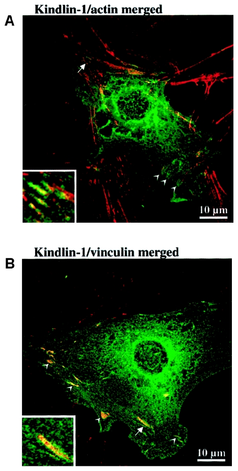
Figure 7.
Intracellular localization of kindlin-1 in epithelial cell line PtK2. A, Merged confocal laser-scanning micrograph of PtK2 cells, transiently transfected with EGFP-kindlin-1 (green) and stained with Alexa Fluor 594 phalloidin to reveal filamentous actin (red). The ends of actin stress fiber bundles exhibit coalignment with EGFP-kindlin-1 to produce the orange coloration in this merged image (arrowheads). These structures were reminiscent of focal contacts. The area marked by the arrow is enlarged in the inset, where the colocalization can be seen more easily. B, Merged confocal laser-scanning micrograph of PtK2 cells transiently transfected with EGFP-kindlin (green) and stained for vinculin by indirect immunofluorescence using Alexa Fluor 594 (red) to reveal focal contacts. Focal contacts (arrowheads) are seen to coalign with EGFP-kindlin to produce the orange coloration in this merged image. Some faint colocalization with cytoskeletal structures also can be seen. The area marked by the arrow is enlarged in the inset, where the colocalization can be seen more easily. These data confirmed that kindlin-1 is a component of focal contacts.

