Abstract
Hereditary leiomyomatosis and renal cell cancer (HLRCC) is an autosomal dominant disorder characterized by smooth-muscle tumors of the skin and uterus and/or renal cancer. Although the identification of germline mutations in the fumarate hydratase (FH) gene in European families supports it as the susceptibility gene for HLRCC, its role in families in North America has not been studied. We screened for germline mutations in FH in 35 families with cutaneous leiomyomas. Sequence analysis revealed mutations in FH in 31 families (89%). Twenty different mutations in FH were identified, of which 18 were novel. Of these 20 mutations, 2 were insertions, 5 were small deletions that caused frameshifts leading to premature truncation of the protein, and 13 were missense mutations. Eleven unrelated families shared a common mutation: R190H. Eighty-one individuals (47 women and 34 men) had cutaneous leiomyomas. Ninety-eight percent (46/47) of women with cutaneous leiomyomas also had uterine leiomyomas. Eighty-nine percent (41/46) of women with cutaneous and uterine leiomyomas had a total hysterectomy, 44% at age ⩽30 years. We identified 13 individuals in 5 families with unilateral and solitary renal tumors. Seven individuals from four families had papillary type II renal cell carcinoma, and another individual from one of these families had collecting duct carcinoma of the kidney. The present study shows that mutations in FH are associated with HLRCC in North America. HLRCC is associated with clinically significant uterine fibroids and aggressive renal tumors. The present study also expands the histologic spectrum of renal tumors and FH mutations associated with HLRCC.
Introduction
Uterine leiomyomas (fibroids) are the most common gynecological tumors in women of reproductive age, with prevalence ranging from 20% to 77% (Cramer and Patel 1990; Stewart 2001). Their presence is the most common indication for hysterectomy in women in the United States (Stewart 2001; Farquhar and Steiner 2002). Historically, the autosomal dominant predisposition to the development of leiomyomas of the skin was referred to as “multiple cutaneous leiomyomatosis” (MIM 150800). Cutaneous leiomyomas are benign smooth-muscle tumors, typically with onset during early adulthood. Cutaneous leiomyomas have been associated with renal carcinomas, uterine leiomyomas, and leiomyosarcomas (Reed et al. 1973; Launonen et al. 2001). In 1973, Reed et al. (1973) described two kindreds in which multiple members, over three generations, exhibited cutaneous leiomyomas, uterine leiomyomas, and/or leiomyosarcomas inherited in an autosomal dominant pattern. The association between cutaneous and uterine leiomyomas is known as “Reed syndrome.”
Recently, Launonen et al. (2001) described a new familial renal cancer syndrome named “hereditary leiomyomatosis and renal cell cancer” (HLRCC [MIM 605839]). They described two kindreds in which cutaneous and uterine leiomyomas and papillary type II renal cell carcinoma segregated together, and, using a genomewide scan, they mapped the HLRCC locus to a region on 1q42-44. Subsequently, an independent group narrowed the leiomyoma locus to a 14-cM region on 1q42.3-43 (Alam et al. 2001). More recently, in collaboration, these groups reported that germline mutations in the FH gene—encoding fumarate hydratase (FH), an enzyme that catalyzes the conversion of fumarate to malate in the Krebs cycle—predispose to dominantly inherited uterine fibroids, skin leiomyoma, and papillary type II renal cell cancer (Tomlinson et al. 2002). These mutations are predicted to result in absent or truncated protein or in substitutions or deletions of highly conserved amino acids. FH most likely acts as a tumor suppressor in HLRCC, since loss of heterozygosity (LOH) studies showed loss of the wild-type allele in cutaneous, uterine, and renal tumors and since the enzyme activity of FH is low or absent in tumors from individuals with leiomyomas (Tomlinson et al. 2002).
Mutations in FH also occur in fumarate hydratase deficiency (FHD [MIM 136850]). Homozygous/compound-heterozygous FH germline mutations cause autosomal recessive FHD, which is characterized by neurological impairment and encephalopathy (Gellera at al. 1990; Bourgeron et al. 1994; Coughlin et al. 1998). Mammalian tissues contain two FH isoenzymes: cytosolic and mitochondrial (Edwards and Hopkinson 1979; Sass et al. 2001). A noticeable exception is the brain, in which only the mitochondrial isoenzymes have been identified (Akiba et al. 1984; Bourgeron et al. 1994). This may explain why FHD predominantly affects brain function.
In 1996, we encountered a family with cutaneous and uterine leiomyomas and papillary renal cancer inherited in an autosomal dominant fashion. The renal tumors had a unique histology, different from classic papillary type I renal cell carcinoma seen in hereditary papillary renal cell carcinoma (HPRC) (Schmidt et al. 1997). In addition, this family’s disease phenotype, now known as “HLRCC,” was not linked to 7q31, and no germline mutations were found in the tyrosine kinase domain of the MET proto-oncogene (unpublished data). To recruit patients in an effort to map and identify the HLRCC-susceptibility gene, we sent two cycles of letters to 11,000 dermatologists throughout the United States and Canada. Similar to previous reports (Alam et al. 2001; Launonen et al. 2001), studies in our laboratory showed that the HLRCC-susceptibility gene maps to 1q42.3, the locus for FH. Subsequently, we screened a cohort of families in North America with HLRCC for mutations in FH. We also recruited patients to investigate the health problems associated with cutaneous leiomyomas, particularly focusing on renal tumors and uterine fibroids. We investigated whether inheritance of germline mutations in FH predisposed individuals in these families to the development of renal tumors or uterine fibroids.
Patients and Methods
Patient Recruitment
We recruited family members with cutaneous leiomyomas by mailing, over a 2-year period, two cycles of ∼22,000 patient-recruitment letters to members of the American Academy of Dermatology. All families were invited to participate in the study regardless of the number of affected individuals in the family and regardless of the presence or absence of associated health problems (renal tumors or uterine fibroids). Subjects were evaluated in consecutive order. An additional three families with HLRCC, which were previously examined in the Clinical Center, were included in the study. The protocol was approved by the institutional review board of the National Cancer Institute. All members of families screened for HLRCC who participated in the present study gave written informed consent.
Patient Evaluation
Families with cutaneous leiomyomas were evaluated at the Clinical Center of the National Institutes of Health and on field trips. Patients were interviewed for a history of cutaneous leiomyomas, uterine fibroids, hysterectomies, and renal tumors. Each patient had a detailed examination (by M.L.T. or J.R.T.) of the skin, including biopsies of selected lesions suspected to be leiomyomas.
For the detection of occult malignancies, family members who came to the National Institutes of Health Clinical Center were examined by computed tomography (CT) scans of the chest, abdomen, and pelvis, followed by renal ultrasound. The kidneys were scanned before and after administration of ∼120 cc Ioxilan 300 (Cook Imaging). High-resolution (1-mm) sections were obtained through the chest, at 10-mm intervals. Renal ultrasound was performed with 3–5-MHz gray-scale and color Doppler transducers with one of two units (Acuson Sequoia or ATL HDI 8000). Women who still had a uterus underwent magnetic resonance imaging (MRI) of the pelvis. Because patients with FHD typically present with severe neurological impairment, we evaluated the present cohort of patients for neurological signs and symptoms.
Definitions
We defined an individual as affected with HLRCC if that individual showed >10 skin lesions clinically compatible with leiomyoma and a minimum of 1 lesion histologically confirmed as a leiomyoma. Histologically, leiomyomas were a proliferation of interlacing bundles of smooth-muscle fibers with a centrally located long blunt-edged nucleus. We defined a family as affected with HLRCC if it contained one or more members affected with cutaneous leiomyomas. Renal tumors were diagnosed on the basis of histologic examination of resected tumors or on the basis of CT scans and renal ultrasound. Renal lesions >1 cm in diameter with >10 Hounsfield units of enhancement that were not cystic on ultrasound were considered to be renal tumors. Uterine fibroids were documented by history, review of medical records, physical examination, MRI, and CT. Hysterectomy was documented by history and a review of medical records, as well as by absence of the uterus on CT scan.
Statistical Analysis
A paired Student’s t test was performed to determine whether there was a difference between the mean age at onset of cutaneous leiomyomas and the mean age at diagnosis of uterine fibroids. All analyses were done on SPSS, version 10.1.4. The possible relationship between renal cancer occurrence and the location of a mutation was assessed using Fisher’s exact test.
Haplotype Analysis
DNA was extracted from peripheral blood or buccal swabs according to standard procedures. Primer sets for microsatellite markers (D1S517, D1S304, D1S180, D1S404, D1S1609, and D1S2836) around the HLRCC locus, at 1q42.3-43, were purchased from Research Genetics. Microsatellite genotyping and haplotype analysis were performed as described elsewhere (Schmidt et al. 2001). Haplotypes were constructed from the genotypes of these six polymorphic microsatellite markers, to identify alleles that were consistently inherited with cutaneous leiomyomas. Haplotypes were generated under the assumption that the smallest numbers of recombinations were present.
Sequencing of the FH Gene
DNA was extracted from peripheral blood leukocytes or buccal swabs according to standard procedures. The genomic sequence that contained FH was determined by BLAST alignment of the mitochondrial FH precursor cDNA (GenBank accession number U59309) with the BAC genomic sequences (GenBank accession numbers AL359764 and AL591898). Primers and PCR conditions are available on request. Supplemental experimental procedures—including methods for identification of exon-intron boundaries, high-throughput DNA sequencing, and analysis—are described in appendix A.
Results
Patient Description
Eighty-one individuals from 35 families exhibited cutaneous leiomyomas. There were 47 women and 34 men, ranging from 22 to 90 years of age. An additional 14 individuals were FH-mutation carriers; of these individuals, the skin status of 6 was unknown, and 8 lacked cutaneous leiomyomas after a detailed skin examination.
Cutaneous Leiomyomas
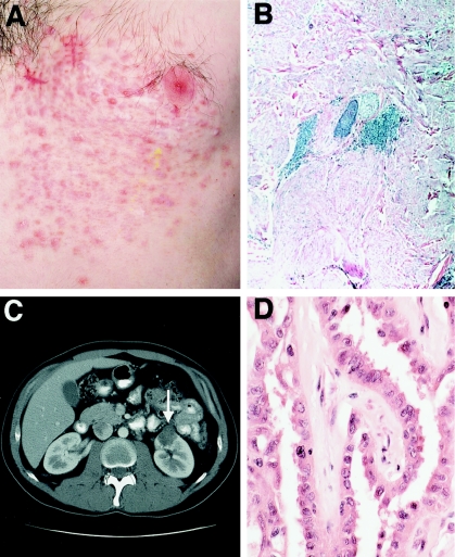
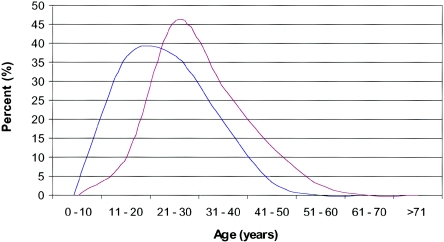
Clinically, cutaneous leiomyomas presented as firm, skin-colored to light-brown-colored papules and nodules (fig. 1). One individual had a cutaneous leiomyosarcoma. The number of lesions ranged from 10 to >100, and the size ranged from 0.4 to 2.5 cm in diameter. An exception was seen in a 22-year-old woman in whom detailed skin examination revealed a single leiomyoma; however, her brother and her father exhibited multiple cutaneous leiomyomas. Patients stated that the number of cutaneous lesions increased with time. Lesions were mostly distributed over the trunk and extremities, except for two patients who also developed multiple lesions on their faces. There were two patterns of cutaneous involvement: disseminated and a combination of disseminated and segmental distribution. Of individuals with cutaneous leiomyomas, 92% complained of pain and paresthesias associated with the cutaneous lesions. As shown in figure 2, the reported onset of cutaneous leiomyomas ranged from 10 to 47 years of age (n=79), with a mean age at onset of 25 years.
Figure 1.
Clinical, histologic, and radiologic manifestations of HLRCC. A, Segmental distribution of leiomyomas in a 32-year-old man. Clinically, cutaneous leiomyomas presented as firm skin-colored to light-brown-colored papules and nodules. B, Histology of cutaneous leiomyoma. Leiomyomas were a proliferation of interlacing bundles of smooth-muscle fibers with a centrally located long blunt-edged nucleus in the dermis. C, Abdominal CT scan, showing the location of a renal tumor in the 32-year-old man shown in panel A. The resected neoplasm was a papillary type II renal cell carcinoma, as shown in panel D. D, Histologic findings of papillary type II renal cell carcinoma. There is a distinct papillary architecture. Cells had an abundant amphophilic cytoplasm and large nuclei with large inclusion-like eosinophilic nucleoli. Magnification × 200.
Figure 2.
Age distribution at diagnosis of uterine fibroids (red) and reported age distribution at onset of cutaneous leiomyomas (blue)
Uterine Fibroids and Related Gynecological Surgeries
Ninety-eight percent (46/47) of women with cutaneous leiomyomas had uterine leiomyomas (fibroids). Uterine fibroids were numerous and large, ranging from 1 to 20 tumors and from 1.5 to 10 cm in diameter, respectively. Patients complained of early onset of fibroid-associated symptoms, including irregular menses, menorrhagia, and pain. Ninety-one percent (42/46) of women with cutaneous and uterine leiomyomas underwent a myomectomy or hysterectomy to treat fibroids. Of these 42 women, 41 underwent a hysterectomy, and 1 had a myomectomy at age 27 years. Fifty-seven percent of women with cutaneous and uterine leiomyomas had a hysterectomy at ⩽30 years of age. Although review of hysterectomy pathology reports did not reveal any uterine leiomyosarcomas, two women had uterine fibroids, with severe atypia in one and mild atypia in the other.
Eight women without a history of a hysterectomy were screened with MRI of the pelvis, for the detection of occult uterine fibroids. Of these eight women, seven had uterine fibroids (five had cutaneous leiomyomas and FH mutations, and two were FH-mutation carriers).
To investigate the clinical presentation of the disease, we plotted the ages at onset of cutaneous leiomyomas and the ages at diagnosis of uterine fibroids. The age at diagnosis of uterine fibroids ranged from 18 to 52 years, with a mean age at diagnosis of 30 years. Cutaneous leiomyomas occurred at a marginally statistically significant (P=.048) earlier mean age (25 years), in comparison with the mean age at diagnosis of uterine fibroids (30 years) (fig. 2).
Uterine fibroids also occurred in 10 women who were FH-mutation carriers. Of these 10 women, 3 did not have cutaneous leiomyomas after a meticulous skin examination, and 7 were deceased (thus, their skin status is unknown). Three other women who were FH-mutation carriers (respectively, 24, 25, and 28 years of age) did not have uterine fibroids after pelvic MRI examination; however, these three women are young and are still at risk for the development of cutaneous and uterine leiomyomas.
Renal Tumors
Thirteen members (five men and eight women) from five families were affected with renal cell carcinoma. In nine cases, pathological confirmation of the diagnosis was obtained; in four cases, the diagnosis of renal carcinoma was based on pathology reports and/or medical records.
Renal carcinomas occurred in eight family members clinically affected with cutaneous leiomyomas and in five carriers or obligate carriers of an FH germline mutation. In addition, we identified three relatives who died of kidney cancer, but no slides or pathology reports were available for review.
Renal tumors were solitary and unilateral. Morphologically, seven renal tumors were classified as papillary type II renal cell carcinoma, one tumor showed features suggestive of collecting duct origin, and another was an unclassified oncocytic tumor. No incipient lesions were identified in the adjacent normal parenchyma. Cases of papillary type II renal cell carcinoma displayed a distinct papillary architecture and characteristic histology (fig. 1). Cells had an abundant amphophilic cytoplasm and large nuclei with large inclusion-like eosinophilic nucleoli. The Furman nuclear grade was 3 or 4 in all cases.
Forty-five individuals with cutaneous leiomyomas had a CT scan of the abdomen and pelvis followed by ultrasound of the kidneys. No evidence of kidney tumors was present in 84.4% (38/45) of individuals. Renal tumors were present in 15.6% (7/45) of individuals (fig. 1). The size of the renal tumors ranged from 3.8 to 14.5 cm in diameter. The median age at detection of renal tumors was 44 years. Four individuals who were diagnosed with renal tumors at ⩽30 years of age (respectively, 23, 26, 28, and 30 years of age) had an aggressive disease. Overall, renal cell carcinoma was associated with an aggressive disease course, with 9 of 13 patients dead of metastatic disease within 5 years of diagnosis.
Other Evaluations
In our clinical evaluation, we considered but did not find the phenotypic expression of two other conditions: FHD, which is an autosomal recessive disease due to mutations in FH, and esophageal leiomyomas. Patients denied neurological symptoms, and a general neurological examination did not reveal gross neurological abnormalities (e.g., microcephaly, hypotonia, and psychomotor retardation). Leiomyomatosis of the esophagus has been reported in other inherited syndromes, including multiple endocrine neoplasia type 1 (McKeeby et al. 2001). The cohort of patients studied denied substernal pain, dysphagia, or other upper-gastrointestinal symptoms typically associated with leiomyomas of the esophagus. Screening CT of the chest in 32 individuals did not show esophageal lesions suggestive of leiomyomas.
Haplotype Analysis and Mutation Analysis
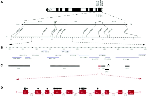
As an initial step in our investigation, we selected three families with HLRCC for the genotyping of six polymorphic microsatellite markers around the leiomyoma locus, at 1q42.3-43 (Alam et al. 2001; Launonen et al. 2001). We constructed haplotypes on the basis of genotyping results, to identify alleles that were consistently inherited with cutaneous leiomyomas. Haplotype analysis consistently showed that a haplotype cosegregated with disease (data not shown). We selected family 214 for linkage analysis, to confirm linkage to 1q42.3-43. A maximum two-point LOD score of 2.47 was obtained for D1S517 at recombination fraction 0 (data not shown). By browsing and searching public and private databases (University of California–Santa Cruz [UCSC] Genome Bioinformatics, the National Center for Biotechnology Information [NCBI], Ensembl Genome Browser, and Celera), we constructed an integrated genetic and physical map of the region of nonrecombination (fig. 3).
Figure 3.
Physical map of the FH critical region on 1q42.3 A, Ideogram of human chromosome 1q long arm with some markers at 1q42.3-43, with integrated genetic map (above) and physical map (below), showing locations of selected polymorphic markers. Cent. = centromere; Tel. = telomere. B, Expanded physical map. The BAC tiling path is shown by blue horizontal lines and GenBank clone ID numbers. BAC overlaps were confirmed in silico. A sequence gap is present in the 2.6-Mb region. C, Locations of known genes, shown within the 2.6-Mb region of FH. Seven known genes were identified and confirmed by searching public and private databases. D, FH exon-intron structure and mutations identified in the exonic sequence.
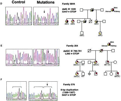
Sequence analysis of the signal peptide, nine coding exons, and splice-site junctions of FH revealed mutations in 89% (31/35) of families with HLRCC (fig. 3 and table 1). We found a total of 20 mutations in the FH coding sequence. Each family's mutation cosegregated with disease and was absent in >210 unaffected individuals. Of these 20 different mutations, 18 were novel, and 2 (R190H and K187R) have previously been reported. Of these 20 mutations, 13 missense mutations were identified; the remaining 7 mutations consisted of 2 insertions and 5 deletions causing frameshifts leading to premature truncation of the protein (fig. 4). No large deletions including the FH gene were identified (data not shown). Eleven unrelated families from diverse ethnic backgrounds and regions had the R190H mutation. Haplotype analysis did not support a founder effect (data not shown). Therefore, the R190H mutation may represent a mutational hotspot. In addition, two unrelated families shared the Y422C mutation.
Table 1.
FH Mutations in Families with HLRCC (N=35)[Note]
| Family | Exon | Mutation | Codon | Predicted Result |
| 260 | 3 | Gdel @ 288 | 97→STOP | Frameshift, protein truncation |
| 8413 | 4 | C431T | S144L | Missense |
| 253 | 4 | A434G | N145S | Missense |
| 8811 | 4 | T455C | M152T | Missense |
| 8411 | 4 | A560G | K187R | Missense |
| 166 | 4 | G569A | R190H | Missense |
| 252 | 4 | G569A | R190H | Missense |
| 262 | 4 | G569A | R190H | Missense |
| 255 | 4 | G569A | R190H | Missense |
| 257 | 4 | G569A | R190H | Missense |
| 258 | 4 | G569A | R190H | Missense |
| 8400 | 4 | G569A | R190H | Missense |
| 8812 | 4 | G569A | R190H | Missense |
| 8425 | 4 | G569A | R190H | Missense |
| 8487 | 4 | G569A | R190H | Missense |
| 8432 | 4 | G569A | R190H | Missense |
| 8401 | 4 | G569T | R190L | Missense |
| 254 | 6 | delGC @ 780 | L260→STOP | Frameshift, protein truncation |
| 8518 | 6 | 7-bp del @ 782–788 | P261→STOP | Frameshift, protein truncation |
| 261 | 6 | C823T | H275Y | Missense |
| 8424 | 6 | T836A | V279D | Missense |
| 8517 | 6 | T875C | L292P | Missense |
| 8423 | 6 | T891A | N297D | Missense |
| 8428 | 6 | G968A | S323N | Missense |
| 8808 | 6 | A964G | S322G | Missense |
| 8810 | 7 | 2-bp ins @ 1004 | E335→STOP | Frameshift, protein truncation |
| 251 | 8 | delA @ 1162 | T388→STOP | Frameshift, protein truncation |
| 259 | 9 | A1265G | Y422C | Missense |
| 8408 | 9 | A1265G | Y422C | Missense |
| 8415 | 9 | 8-bp dup @ 1300–1307 | S437→STOP | Frameshift, protein truncation |
| 6844 | 9 | delG @ 1339 | G447→STOP | Frameshift, protein truncation |
| 700 | ND | ND | ND | ND |
| 572 | ND | ND | ND | ND |
| 501 | ND | ND | ND | ND |
| 511 | ND | ND | ND | ND |
Note.— Mutations are named according to the recommendations of the Nomenclature System for Human Gene Mutations. Nucleotide numbering is according to the cytosolic FH sequence (GenBank accession number NM_000143), with the A of the ATG initiator codon as nucleotide position 1. Amino acid positions were numbered from the translation of the cytosolic FH nucleotide sequence. ND = not detected.
Figure 4.
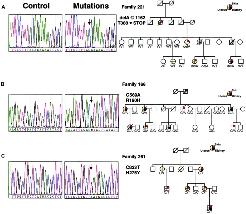
FH mutations in patients with HLRCC. Sequencing chromatograms of genomic DNA from control subjects and patients are shown at left (arrows indicate the position of the identified nucleotide changes), and pedigrees are shown at right (asterisks indicate history of renal cancer). The delA at nucleotide 1162 (A), a delG at nucleotide 1339 (D), a delGC at nucleotides 780 and 781 (E), and an 8-bp duplication at nucleotides 1300–1307 (F) cause shifts in the reading frame that lead to a stop codon downstream and truncate the corresponding protein; a missense mutation, G569A (B), changes an arginine to a histidine at codon 190, and another missense mutation, C823T (C), changes a histidine to a tyrosine at codon 275. Sequences containing insertions and deletions are derived from subcloned alleles from affected patients.
Genotype-Phenotype Correlation
We found mutations in all exons except 1, 2, and 5. Exon 4 (in 16 families with mutations) and exon 6 (in 8 families with mutations) had the majority of the mutations (fig. 3). Tomlinson et al. (2002) reported that the location of FH mutations associated with FHD differs significantly from that associated with leiomyomatosis. They found that mutations in families with leiomyomatosis occurred toward the amino terminus of FH, with 22 of 24 located before codon 250, whereas 8 of the 10 mutations reported to be associated with FHD were located after codon 250 (P<.0001). Therefore, a comparison of the reported FH mutations associated with FHD and the FH mutations identified in the present study was conducted. We found that 17 of 31 FH mutations in the families studied are located before codon 250. In contrast, 8 of the 10 FH mutations reported in FHD were located after codon 250 (P=.075).
We also compared the location of FH mutations found in patients with and without renal cancer. Whereas one family with renal cancer had a mutation before codon 250, four families with renal cancer had mutations after codon 250 (P=.148). Two families with renal cancer had missense mutations (H275Y and R190L), and three families with renal cancer had frameshift mutations (an AA insertion at nucleotide position 1004, resulting in a stop codon 2 amino acids downstream; an A deletion at nucleotide position 1162, resulting in a stop codon 16 amino acids downstream; and a G deletion at nucleotide position 1339, resulting in a stop codon 10 amino acids downstream) (fig. 4). Four of these five mutations were associated with papillary type II renal cell carcinoma. The G deletion at nucleotide position 1339 was identified in an individual affected with collecting duct carcinoma (CDC) of the kidney.
All the families with HLRCC except one (family 8401) included women affected with uterine fibroids. There was no correlation between the location or type of mutation and uterine fibroids. In addition, there was no correlation between the type of mutation and the severity of disease in terms of onset, survival, or number of individuals affected (data not shown).
Discussion
We characterized the clinical and genetic features of 35 families with HLRCC in North America. In 89% (31/35) of the cohort of families studied in North America, sequence analysis revealed germline mutations in FH. In contrast, the Leiomyoma Consortium identified germline mutations in FH in 60% (25/42) of the cohort of European families with leiomyomatosis that they studied. We did not identify mutations in four of the families that we studied. This may be due to either sequence changes in introns that affect splicing or single-nucleotide substitutions in the 3′ UTR of FH. We did not find large deletions of the entire FH gene in the cohort of individuals studied (data not shown). Large deletions including the entire FH gene, of ∼2.4 and ∼1.9 Mb, were identified in two English families by use of FISH (Tomlinson et al. 2002). We identified 20 different FH mutations, of which 18 were novel. Of these 20 mutations, 7 were frameshifts (2 insertions and 5 deletions) leading to premature truncation of the protein, and 13 were missense changes predicted to result in substitution of the amino acids.
Of the families with HLRCC that we studied, 55% (17/31) had FH mutations before codon 250. In contrast, the Leiomyoma Consortium found that 92% (22/24) of mutations associated with HLRCC were located before codon 250 (Tomlinson et al. 2002). As we continue to identify additional FH mutations, we expect to confirm our finding that FH mutations associated with HLRCC are distributed throughout the gene, rather than mostly localized to the amino-terminal half of FH.
The distinction between the phenotypic expression of HLRCC and that of FHD is most likely due to the pattern of inheritance, rather than to the location of the FH mutation. FHD arises when there is an autosomal recessive inheritance that results in germline biallelic mutations in FH, and it is characterized by severe neurological impairment, including hypotonia, seizures, and cerebral atrophy. The deprivation of aerobic energy due to loss of function of the FH mitochondrial isoenzyme in brain tissue at early embryogenesis may interfere with normal fetal brain development. This may explain the neurological impairment associated with FHD. Leiomyomas and renal cancer have not been reported in individuals affected with FHD; however, most individuals with FHD survive only a few months, and very few survive early adulthood. Parents (FH-mutation heterozygous) of individuals affected with FHD have been reported to develop cutaneous leiomyomas, as present in individuals affected with HLRCC (Tomlinson et al. 2002). In contrast to FHD, for HLRCC, germline mutations in FH are inherited in an autosomal dominant pattern, leading to predisposition to the development of uterine and cutaneous leiomyomas and renal cancer. We also found that the cohort of patients with HLRCC that we studied did not develop hypotonia, seizures, or neurological impairment. This evidence, in combination with the distribution of FH mutations associated with HLRCC throughout the gene, strongly suggests that the pattern of inheritance of FH mutations determines the phenotypic expression of disease.
We identified renal cancer in 5 of 35 families. Histologically, papillary renal cell carcinoma constitutes 15% of all renal cancer (Kovacs et al. 1997). Papillary type II renal cell carcinoma, an uncommon histologic type of renal cell cancer, was the predominant type of renal tumor that occurred in the patients whom we studied. To our knowledge, only three families with HLRCC have been described in the renal cancer literature (Kiuru et al. 2001; Launonen et al. 2001). Germline mutations in FH have been reported in these three Finnish kindreds with papillary type II renal cell carcinoma. Two families shared a 2-base deletion in codon 181, and the other had the Arg300X mutation (Tomlinson et al. 2002). The failure to identify renal tumors in families with leiomyomatosis described previously (Alam et al. 2001) may be due, in part, to the lack of screening for renal tumors or to the possibility that papillary tumors can be missed by renal ultrasound (Choyke et al. 1997). In the present study, we also describe the first reported case of HLRCC suggestive of CDC of the kidney, with germline mutation in FH. Interestingly, this is consistent with reports, in the literature, of cytogenetic abnormalities found in CDC, including monosomy of chromosome 1, as well as LOH of 1q (reported to occur in 60%–80% of cases) (Steiner et al. 1996; Fuzesi et al. 1992). Furthermore, the patient whom we studied had the typical clinical presentation of CDC, which is characterized by an aggressive disease course with evidence of metastatic disease at the time of presentation and poor prognosis (Kennedy et al. 1990). Therefore, the present study expands the histologic spectrum of renal tumors in HLRCC.
The biologic characteristics of the renal tumors also provided support for the concept that HLRCC conferred an inherited predisposition to the development of renal tumors. Absence or low FH enzyme activity and loss of the wild-type allele in renal tumors suggest that FH is involved in the development of renal tumors (Tomlinson et al. 2002). However, several lines of evidence suggest that modifying genes, environmental factors, or other tumorigenic mechanisms may play a role in the development of renal tumors. First, renal tumors in HLRCC are solitary and unilateral, in contrast to renal tumors in VHL, HPRC, and BHD, which are usually multiple and bilateral and for which numerous microscopic renal-tumor precursors have been observed within the normal renal parenchyma (Walther et al. 1995; Ornstein et al. 2000; Zbar et al. 2002); furthermore, the lack of multifocal disease in the kidney and the presence of segmental distribution of skin lesions suggest the possibility of mosaicism in some affected individuals with HLRCC. Second, whereas only 10%–15% of families with HLRCC develop kidney cancer, 80%–90% of families with VHL and HPRC have one or more individual with kidney cancer. Third, there was variability of expression of renal tumors both between and within families; family 214 had the R190H mutation and included four individuals affected with renal tumors, whereas an additional 10 families who shared the R190H mutation and were screened for occult renal tumors lacked individuals affected with renal tumors. These findings suggest that the existence of other genetic and/or environmental factors may influence the phenotype.
The cohort of women with cutaneous leiomyomas that we studied had a higher prevalence and an earlier age at diagnosis of uterine fibroids than did women in the general population. Ninety-eight percent had uterine fibroids, with a mean age at diagnosis of 30 years. In the general population, the reported prevalence rates of uterine fibroids range from 22% to 77%, with the highest prevalence in women 40–44 years of age (Marshall et al. 1997; Stewart et al. 2001). In addition, 89% of women in the cohort of women with cutaneous leiomyomas that we studied had a hysterectomy for symptomatic uterine fibroids. Of these, 57% had a hysterectomy at ⩽30 years of age. In contrast, hysterectomy surveillance in the United States from 1994 to 1999 showed that hysterectomy was most frequent for those 40–44 years of age, with 52% of women undergoing hysterectomy at ⩽44 years of age (Keshavarz et al. 2002). The early age at onset of symptomatic uterine fibroids had a significant impact on the childbearing years of women with HLRCC.
In conclusion, germline mutations in FH predispose families in North America to the development of cutaneous and uterine leiomyomas and renal cancer. The mechanisms by which FH defects promote tumorigenesis are unknown, but may involve alterations in citrate production, activation of hypoxia pathways (including HIF-1), or free radical formation. In addition to cutaneous leiomyomas, HLRCC is associated with clinically significant uterine fibroids and aggressive renal tumors. Screening for renal tumors is best done with CT scans of the abdomen and pelvis. Renal ultrasounds alone are not sufficient, because papillary renal tumors are often isoechoic and can be missed on ultrasound. A characteristic feature of many papillary tumors is a lesion that is seen on CT but may not be detected on ultrasound examination. HLRCC needs to be considered in the differential diagnosis in any patient with renal cancer, ranging from papillary type II renal cell carcinomas to renal tumors with collecting duct histology that are unilateral and solitary. It also needs to be considered in the differential diagnosis of women with a family history of renal cancer and early onset of multiple uterine fibroids. Appropriate counseling is necessary for women, to insure informed reproductive decisions.
Acknowledgments
We thank the members of the American Academy of Dermatology, for their help in the recruitment of families; the families, for their participation in the study; Dr. Lifang Hou, for her technical assistance; Dr. Mahul Amin and Dr. Victor Reuter, for consultations on renal-tumor pathology; and Birgitta Sievers, Cia Manolatos, Robin Eyler, Kathleen Hurley, James Peterson, and Lindsay Middelton, for their many contributions to this project. The content of this publication does not necessarily reflect the views or policies of the Department of Health and Human Services, nor does mention of trade names, commercial products, or organizations imply endorsement by the U.S. government. This project has been funded in part with federal funds from the National Cancer Institute, National Institutes of Health, under contract N01-CO-12400.
Appendix A: Supplementary Data
Two-point LOD scores were calculated with Mlink in Linkage package, version 5.1 (Lathrop et al. 1984), using the high-penetrance model. Autosomal dominant inheritance was assumed, with a gene frequency of 0.0001 and three liability classes. Allele frequencies were estimated on the basis of the genotypes of unrelated spouses. Individuals were considered as affected if they had cutaneous leiomyomas. The genomic and BAC sequences were derived from available public and private databases (UCSC Genome Bioinformatics, NCBI, Ensembl Genome Browser, and Celera). The locations of BAC sequences and the genetic markers on the physical map were determined by the BLAST 2 (Tatusova and Madden 1999; also see the NCBI BLAST Home Page) and BLAT (Kent 2002; also see the UCSC Genome Bioinformatics Web site) algorithms. A 6.2-Mb region of the assembled genomic sequence was extracted by using a distal marker, D1S517, and a proximal marker, D1S423. The known and predicted genes were identified by browsing and searching databases (UCSC Genome Bioinformatics, NCBI, Ensembl Genome Browser, and Celera). Exon-intron structure of FH was determined by analysis of the genomic structure, using the Wise2 program. An upstream mitochondrial signal peptide (termed “exon 0”) was localized by Signal P (Nielsen et al. 1997). In addition, nine coding exons were identified. Amplicons were designed to evaluate coding sequences and splice junctions. Primers located in neighboring introns ⩾20 bp from the splice junctions were designed with the aid of Oligo Tech, version 1 (Oligos Etc & Oligo Therapeutics). Standard PCR conditions were employed with AmpliTaq (Perkin Elmer) or Taq polymerases (Invitrogen). PCR components are standard. PCR products were quantitated by agarose gel electrophoresis and were purified using Multiscreen PCR cleanup plates (Millipore).
Double-stranded sequencing reactions (10 μl) using Big Dye Terminators ready reaction mix (Applied Biosystems) were purified using Performa plates (Edge Biosystems) and were electrophoresed on an ABI 3700 genetic analyzer. Chromatograms were aligned and analyzed using Lasergene (DNAStar). Alignments were examined using the conflict finder, to locate Phred-identified discrepancies; then, forward and reverse chromatograms from each affected patient were manually examined, to locate additional secondary peaks. Sequence variants found in one or more affected patients (but not in unaffected individuals) were examined for cosegregation with disease in their respective families by denaturing high-performance liquid chromatography (DHPLC) or single-stranded sequencing. Insertions and deletions were subcloned with a Topo Cloning Kit (Invitrogen) and were sequenced. A minimum of 210 normal individuals (420 chromosomes) were examined for the presence of each disease-associated sequence variant. DHPLC was performed using a Transgenomic Wave system with a DNASep column. Temperature predictions were obtained by the Stanford Melt algorithm (see the DHPLC Melt Program Web site) or Wavemaker (Transgenomic).
Electronic-Database Information
Accession numbers and URLs for data presented herein are as follows:
- Celera, http://www.celera.com/
- DHPLC Melt Program, http://insertion.stanford.edu/melt.html
- Ensembl Genome Browser, http://www.ensembl.org/
- GenBank, http://www.ncbi.nlm.nih.gov/Genbank/ (for mitochondrial FH precursor cDNA [accession number U59309] and BAC genomic sequences [accession numbers AL359764 and AL591898])
- National Center for Biotechnology Information, http://www.ncbi.nlm.nih.gov/
- NCBI Blast Home Page, http://www.ncbi.nlm.nih.gov/BLAST/ (for BLAST 2)
- Online Mendelian Inheritance in Man (OMIM), http://www.ncbi.nlm.nih.gov/Omim/ (for multiple cutaneous leiomyomatosis, HLRCC, and FHD)
- UCSC Genome Bioinformatics, http://genome.ucsc.edu/ (for Genome Browser and BLAT)
- Wise2, http://www.ebi.ac.uk/Wise2/
References
- Akiba T, Hiraga K, Tuboi S (1984) Intracellular distribution of fumarase in various animals. J Biochem 96:189–195 [DOI] [PubMed] [Google Scholar]
- Alam NA, Bevan S, Churchman M, Barclay E, Barker K, Jaeger EE, Nelson HM, Healy E, Pembroke AC, Friedmann PS, Dalziel K, Calonje E, Anderson J, August PJ, Davies MG, Felix R, Munro CS, Murdoch M, Rendall J, Kennedy S, Leigh IM, Kelsell DP, Tomlinson IP, Houlston RS (2001) Localization of a gene (MCUL1) for multiple cutaneous leiomyomata and uterine fibroids to chromosome 1q42.3-q43. Am J Hum Genet 68:1264–1269 [DOI] [PMC free article] [PubMed] [Google Scholar]
- Bourgeron T, Chretien D, Poggi-Bach J, Doonan S, Rabier D, Letouze P, Munnich A, Rotig A, Landrieu P, Rustin P (1994) Mutation of the fumarase gene in two siblings with progressive encephalopathy and fumarase deficiency. J Clin Invest 93:2514–2518 [DOI] [PMC free article] [PubMed] [Google Scholar]
- Choyke PL, Walther MM, Glenn GM, Wagner JR, Venzon DJ, Lubensky IA, Zbar B, Linehan WM (1997) Imaging features of hereditary papillary renal cancers. J Comput Assist Tomogr 21:737–741 [DOI] [PubMed] [Google Scholar]
- Coughlin EM, Christensen E, Kunz PL, Krishnamoorthy KS, Walker V, Dennis NR, Chalmers RA, Elpeleg ON, Whelan D, Pollitt RJ, Ramesh V, Mandell R, Shih VE (1998) Molecular analysis and prenatal diagnosis of human fumarase deficiency. Mol Genet Metab 63:254–262 [DOI] [PubMed] [Google Scholar]
- Cramer SF, Patel A (1990) The frequency of uterine leiomyomas. Am J Clin Pathol 94:435–438 [DOI] [PubMed] [Google Scholar]
- Edwards YH, Hopkinson DA (1979) The genetic determination of fumarase isozymes in human tissues. Ann Hum Genet 42:303–313 [DOI] [PubMed] [Google Scholar]
- Farquhar CM, Steiner CA (2002) Hysterectomy rates in the United States 1990–1997. Obstet Gynecol 99:229–234 [DOI] [PubMed] [Google Scholar]
- Fuzesi L, Cober M, Mittermayer C (1992) Collecting duct carcinoma: cytogenetic characterization. Histopathology 21:155–160 [DOI] [PubMed] [Google Scholar]
- Gellera C, Uziel G, Rimoldi M, Zeviani M, Laverda A, Carrara F, DiDonato S (1990) Fumarase deficiency is an autosomal recessive encephalopathy affecting both the mitochondrial and the cytosolic enzymes. Neurology 40:495–499 [DOI] [PubMed] [Google Scholar]
- Kennedy SM, Merino MJ, Linehan WM, Roberts JR, Robertson CN, Neumann RD (1990) Collecting duct carcinoma of the kidney. Hum Pathol 21:449–456 [DOI] [PubMed] [Google Scholar]
- Kent WJ (2002) BLAT—the BLAST-like alignment tool. Genome Res 12:656–664 [DOI] [PMC free article] [PubMed] [Google Scholar]
- Keshavarz H, Hillis SD, Kieke BA, Marchbanks PA (2002) Hysterectomy surveillance—United States, 1994–1999. MMWR Morb Mortal Wkly Rep 51(SS05):1–8 [Google Scholar]
- Kiuru M, Launonen V, Hietala M, Aittomaki K, Vierimaa O, Salovaara R, Arola J, Pukkala E, Sistonen P, Herva R, Aaltonen LA (2001) Familial cutaneous leiomyomatosis is a two-hit condition associated with renal cell cancer of characteristic histopathology. Am J Pathol 159:825–829 [DOI] [PMC free article] [PubMed] [Google Scholar]
- Kovacs G, Akhtar M, Beckwith BJ, Bugert P, Cooper CS, Delahunt B, Eble JN, Fleming S, Ljungberg B, Medeiros LJ, Moch H, Reuter VE, Ritz E, Roos G, Schmidt D, Srigley JR, Störkel S, van den Berg E, Zbar B (1997) The Heidelberg classification of renal cell tumors. J Pathol 183:131–133 [DOI] [PubMed] [Google Scholar]
- Lathrop GM, Lalouel JM, Julier C, Ott J (1984) Strategies for multilocus linkage analysis in humans. Proc Natl Acad Sci USA 81:3443–3446 [DOI] [PMC free article] [PubMed] [Google Scholar]
- Launonen V, Vierimaa O, Kiuru M, Isola J, Roth S, Pukkala E, Sistonen P, Herva R, Aaltonen LA (2001) Inherited susceptibility to uterine leiomyomas and renal cell cancer. Proc Natl Acad Sci USA 98:3387–3392 [DOI] [PMC free article] [PubMed] [Google Scholar]
- Marshall LM, Spiegelman D, Barbieri RL, Goldman MB, Manson JE, Colditz GA, Willett WC, Hunter DJ (1997) Variation in the incidence of uterine leiomyomata among premenopausal women by age and race. Obstet Gynecol 90:967–973 [DOI] [PubMed] [Google Scholar]
- McKeeby JL, Li X, Zhuang Z, Vortmeyer AO, Huang S, Pirner M, Skarulis MC, James-Newton L, Marx SJ, Lubensky IA (2001) Multiple leiomyomas of the esophagus, lung, and uterus in multiple endocrine neoplasia type 1. Am J Pathol 159:1121–1127 [DOI] [PMC free article] [PubMed] [Google Scholar]
- Nielsen H, Engelbrecht J, Brunak S, von Heijne G (1997) Identification of prokaryotic and eukaryotic signal peptides and prediction of their cleavage sites. Protein Engineering 10:1–6 [DOI] [PubMed] [Google Scholar]
- Ornstein DK, Lubensky IA, Venzon D, Zbar B, Linehan WM, Walther MM (2000) Prevalence of microscopic tumors in normal appearing renal parenchyma of patients with hereditary papillary renal cancer. J Urol 163:431–433 [PubMed] [Google Scholar]
- Reed WB, Walker R, Horowitz R (1973) Cutaneous leiomyomata with uterine leiomyomata. Acta Derm Venereol 53:409–416 [PubMed] [Google Scholar]
- Sass E, Blachinsky E, Karniely S, Pines O (2001) Mitochondrial and cytosolic isoforms of yeast fumarase are derivatives of a single translation product and have identical amino termini. J Biol Chem 276:46111–46117 [DOI] [PubMed] [Google Scholar]
- Schmidt L, Duh FM, Chen F, Kishida T, Glenn G, Choyke P, Scherer S, et al (1997) Germline and somatic mutations in the tyrosine kinase domain of the MET proto-oncogene in papillary renal carcinomas. Nat Genet 16:68–73 [DOI] [PubMed] [Google Scholar]
- Schmidt LS, Warren MB, Nickerson ML, Weirich G, Matrosova V, Toro JR, Turner ML, Duray P, Merino M, Hewitt S, Pavlovich CP, Glenn G, Greenberg CR, Linehan WM, Zbar B (2001) Birt-Hogg-Dubé syndrome, a genodermatosis associated with spontaneous pneumothorax and kidney neoplasia, maps to chromosome 17p11.2. Am J Hum Genet 69:876–882 [DOI] [PMC free article] [PubMed] [Google Scholar]
- Steiner G, Cairns P, Polascik TJ, Marshall FF, Epstein JI, Sidransky D, Schoenberg M (1996) High-density mapping of chromosomal arm 1q in renal collecting duct carcinoma: region of minimal deletion at 1q32.1-32.2. Cancer Res 56:5044–5046 [PubMed] [Google Scholar]
- Stewart EA (2001) Uterine fibroids. Lancet 357:293–298 [DOI] [PubMed] [Google Scholar]
- Tatusova TA, Madden TL (1999) BLAST 2 sequences, a new tool for comparing protein and nucleotide sequences. FEMS Microbiol Lett 174:247–250 (erratum 177:187–188) [DOI] [PubMed] [Google Scholar]
- Tomlinson IP, Alam NA, Rowan AJ, Barclay E, Jaeger EE, Kelsell D, Leigh I, et al (2002) Germline mutations in FH predispose to dominantly inherited uterine fibroids, skin leiomyomata and papillary renal cell cancer. Nat Genet 30:406–410 [DOI] [PubMed] [Google Scholar]
- Walther M M, Lubensky IA, Venzon D, Zbar B, Linehan WM (1995) Prevalence of microscopic lesions in grossly normal renal parenchyma from patients with von Hippel–Lindau disease, sporadic renal cell carcinoma and no renal disease: clinical implications. J Urol 154:2010–2014 [PubMed] [Google Scholar]
- Zbar B, Alvord WG, Glenn G, Turner M, Pavlovich CP, Schmidt L, Walther M, Choyke P, Weirich G, Hewitt SM, Duray P, Gabril F, Greenberg C, Merino MJ, Toro J, Linehan WM (2002) Risk of renal and colonic neoplasms and spontaneous pneumothorax in the Birt-Hogg-Dube syndrome. Cancer Epidemiol Biomarkers Prev 11:393–400 [PubMed] [Google Scholar]


