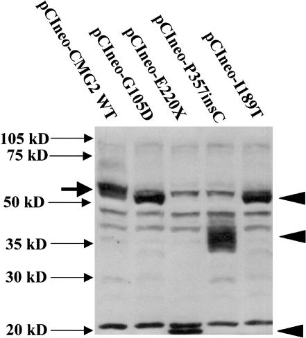
Figure 5.
CMG-2 mutations, resulting in altered CMG-2 protein expression, as detected by western blotting. Two hundred ninety-three cells were transfected with 1.5 mg of plasmid DNA (in six-well dishes) with Lipofectamine 2000 and various CMG-2 WT and mutant constructs, as indicated. Following transfection, cells were lysed after 24 h with 0.5 ml SDS-PAGE sample buffer, containing mercaptoethanol, and were treated at 100°C for 10 min. Thirty milliliters of sample was loaded per lane on a 10% SDS-PAGE gel, and protein samples were transferred to PVDF membranes and were probed with anti-CMG-2 affinity-purified antibodies (1 mg/ml), as described elsewhere (Bell et al. 2001). Closed arrowheads indicate the position of anti-CMG-2 reactive mutant proteins. Solid arrow indicates the position of CMG-2 WT protein observed in 293 cells transfected with pCIneo-CMG2-WT.

