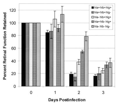
FIG. 3.
Contribution of specific membrane-damaging toxins to decreases in retinal function during experimental S. aureus endophthalmitis. Approximately 100 CFU of the following S. aureus strains was injected into the vitreous at day 0 (16, 17): hla+hlb+hlg+, wild-type strain; hla+hlb+hlg−, isogenic mutant lacking gamma-hemolysin; hla−hlb+hlg+, isogenic mutant lacking alpha-toxin; hla+hlb−hlg+, isogenic mutant lacking beta-toxin; and hla−hlb−hlg−, isogenic mutant lacking alpha-toxin, beta-toxin, and gamma-hemolysin (strains provided by Tim Foster, Trinity College). Data at each postinfection day are presented as percentages of the B-wave amplitude of retinal function retained compared to baseline and sham-injected controls. Electroretinography losses were averaged for each group and compared using the Student t test. Values and error bars represent means ± standard errors of the means for four or more eyes per group.

