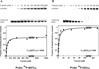
Figure 5.
Gel mobility shift assays of the binding of Translin to the single-stranded oligodeoxynucleotide d(GT)15 and the single-stranded oligoribonucleotide (GU)15. Recombinant S.pombe Translin at the indicated concentrations was incubated with each of the two single-stranded 32P-labeled oligonucleotides. Complexes were analyzed by the gel mobility shift technique. The phosphorimager images of the gels are shown at the top of (A) and (B). Plots of the fraction of labeled oligonucleotide in the complex versus the total concentration of Translin in the binding reaction are shown at the bottom. (A) Binding of the S.pombe Translin to the oligodeoxynucleotide d(GT)15 at a concentration of 0.04 nM. (B) Binding of the S.pombe Translin to the oligoribonucleotide (GU)15 at a concentration of 0.03 nM. Kdis values were derived from these plots, as described previously (5).

