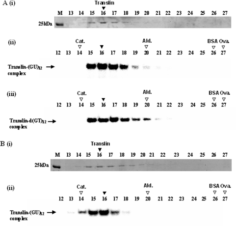
Figure 8.
Glycerol gradient centrifugation of the recombinant S.pombe Translin. (A) Sample of the recombinant S.pombe Translin was centrifuged in a 20–40% linear glycerol gradient, as described in Materials and Methods. Fractions of equal volumes were collected from the bottom of the gradient. Aliquots withdrawn from these fractions were analyzed by SDS–gel electrophoresis, followed by Commassie blue staining [A (i)]. Other aliquots withdrawn from the same fractions were analyzed by the gel mobility shift assay for binding the 32P-labeled oligoribonucleotide (GU)12 [A (ii)] or the 32P-labeled oligodeoxynucleotide d(GT)12 [A (iii)]. Another sample of the recombinant S.pombe Translin was preincubated with the 32P-labeled oligoribonucleotide (GU)12 and then centrifuged in another tube in the same run. Aliquots withdrawn from this tube were also analyzed by SDS–gel electrophoresis and Coomassie blue staining [B (i)]. Other aliquots were electrophoresed in a native polyacrylamide gel and the 32P-labeled bands were detected by phosphorimaging [B (ii)]. M indicates the gel lanes including the gel marker proteins. Horizontal arrows indicate the gel-shifted complexes generated by binding of Translin. Vertical filled arrowheads indicate the peaks of the Coomassie blue-stained Translin, and the peaks of the RNA- and DNA-binding activities. Vertical open arrowheads indicate the peaks of the centrifugation marker proteins: Cat., Catalase (232 kDa); Ald., Aldolase (158 kDa); BSA (66 kDa); Ova., Ovalbumin (43 kDa).

