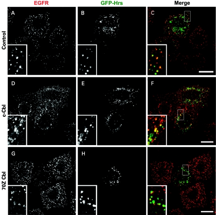
Figure 2. Co-localization of EGFR and GFP–Hrs in cells expressing c-Cbl and 70Z-Cbl mutant.
HeLa cells were seeded on to coverslips and transfected with GFP–Hrs and pCDNA3 (control), HA-tagged c-Cbl or 70Z-Cbl and then stimulated for 8 min with 100 ng/ml EGF 22 h post-transfection after a 16 h starvation period. The cells were fixed with 3% paraformaldehyde, permeabilized with Triton X-100 and stained with anti-EGFR (EGFR shown in red) followed by a secondary antibody coupled with Alexa Fluor 594. All panels show a single confocal section. Scale bars, 20 μm.

