Figure 5.
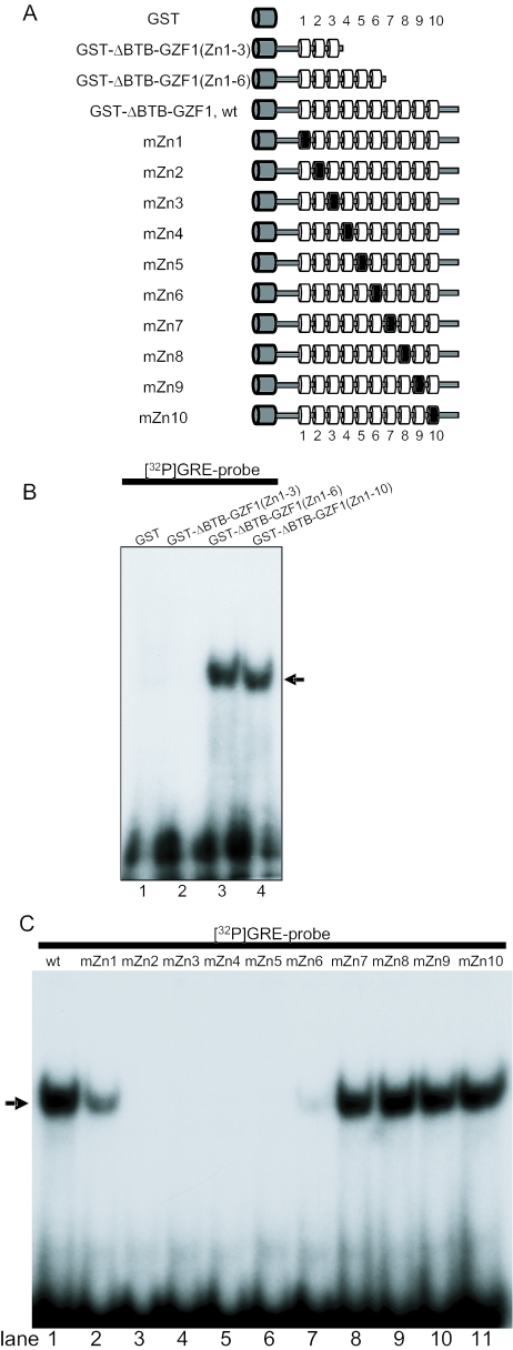
Analysis of zinc finger motifs in GZF1 required for DNA binding. (A) Schematic representation of GZF1 zinc finger motif mutants used for EMSA. Two deletion zinc finger mutants are shown. Two cysteine residues in the C2H2 motifs of each finger were replaced with arginines. The wild-type and mutated fingers are shown as a white and a black cylinder, respectively. GST is shown as a grey cylinder. (B) Two deletion mutants of zinc finger motifs in GZF1 were subjected to EMSA. The arrow indicates the DNA–protein complex. (C) EMSA with GZF1 constructs mutated in the individual zinc finger motifs.

