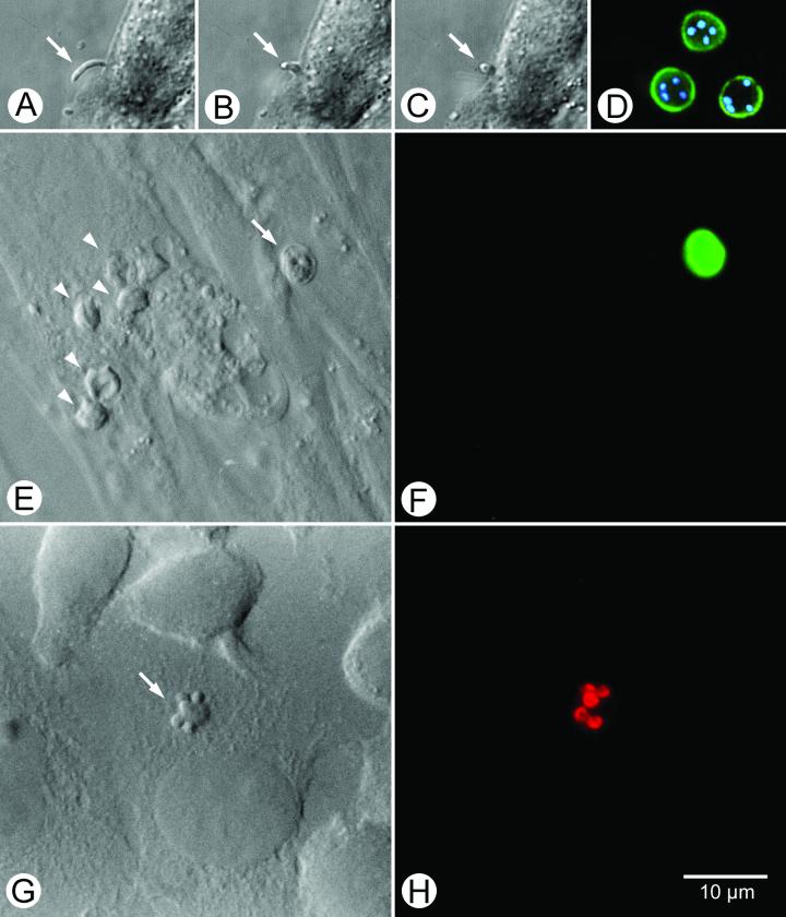
FIG. 3.
Panels A, B, and C show the progressive invasion of a host cell by a sporozoite of C. parvum. Panel D shows fluorescein-conjugated monoclonal antibody-labeled oocysts with internal sporozoite nuclei stained with the dye DAPI. Panel E shows the differential interference contrast bright-field view of host cells with developing asexual parasite life cycle stages (arrowheads) and an oocyst (arrow). The latter is part of the inoculum, as revealed by the bright green label evident in panel F, a result of the FTSC labeling process. Panel G shows a differential interference contrast view of a type I meront with immature (“budding”) merozoites (arrow) labeled with a monoclonal antibody conjugated with the fluorescent dye indocarbocyanine.

