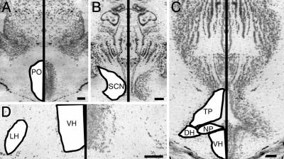
Fig. 2.
Photomicrographs of cresyl-violet-stained transverse sections through frog brain. Left half of photographs are mirror images of right half for clarity. Hypothalamic nuclei are labeled on figures based on standard nomenclature (31) with the following abbreviations: DH, dorsal hypothalamus; NP, nucleus of the periventricular organ; POA, anterior POA; VH, ventral hypothalamus. (Bar, 100 μm.) (A) The second-most rostral section we used for POA analysis. (B) Section containing the SCN comparable to figure 3A in ref. 31. (C) Third-most rostral section containing the infundibular hypothalamus, equivalent to figure 5A in ref. 31. (D) Higher-magnification image showing sparse cells in the LH at a level corresponding to figure 4A of ref. 31.

