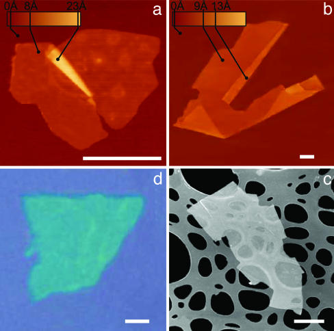
Fig. 1.
2D crystal matter. Single-layer crystallites of NbSe2 (a), graphite (b), Bi2Sr2CaCu2Ox (c), and MoS2 (d) visualized by AFM (a and b), by scanning electron microscopy (c), and in an optical microscope (d). (All scale bars: 1 μm.) The 2D crystallites are on top of an oxidized Si wafer (300 nm of thermal SiO2) (a, b, and d) and on top of a holey carbon film (c). Note that 2D crystallites were often raised by an extra few angstroms above the supporting surface, probably because of a layer of absorbed water. In such cases, the pleated and folded regions seen on many AFM images and having the differential height matching the interlayer distance in the corresponding 3D crystals help to distinguish between double-layer crystals and true single sheets such as those shown here.

