
Figure 4.
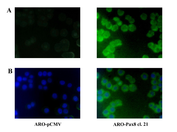
Immunofluorescent localization of NIS in ARO cells. ARO-pCMV and ARO-Pax8 cl. 21 cells were analyzed by immunofluorescence microscopy using rabbit polyclonal anti-NIS and anti-rabbit FITC secondary antibody. A: NIS (green) localization; B. Double immunofluorescence with blue that indicates DAPI nuclear staining and green that indicates the NIS localization. Magnification: 40×.