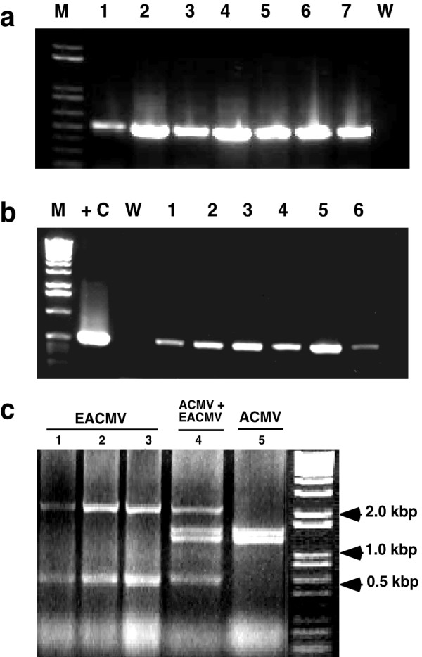
Figure 4.

Analysis of geminivirus DNA eluted from FTA-preserved leaf tissues of infected maize and cassava plants growing in farmer's fields in Kenya and Malawi. (a) detection of maize streak virus from infected plants in Malawi. Primers MSV-F and MSV-R (Table 1) were used to amplify a 500 bp fragment from the conserved region of this monopartite geminivirus. (b) detection of East African cassava mosaic virus-like sequences from leaf tissues pressed onto FTA cards plants in Malawi. Primers EAB555F and EAB555R (Table 1) were used to amplify a 550 bp fragment of the B genomic component. (c) Restriction analysis of whole A genomic components (2.8 kb) of East African cassava mosaic virus (EACMV) and African cassava mosaic virus (ACMV) isolated from FTA leaf presses of diseased cassava leaves sampled in Western Kenya. The amplified PCR product was cloned into pGEM-T Easy vector (Promega), the DNA amplified by miniprep and digested with EcoRV for 1.5 hrs at 37°C. Unique bands generated by this restriction enzyme facilitate identification of single infections with EACMV and ACMV (lanes 1–3 and 5 respectively) and a plant co-infected with both geminivirus species (lane 4). M: marker, +C: positive control, W: water control.
