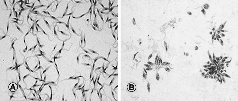
FIG. 2.
(A) Epimastigote stage of T. cruzi from culture, the stage normally seen in the insect vector. Flagella can be seen arising from the slender organisms. The dark-staining body within the cell is the nucleus. Magnification, ×1,000. (B) Promastigote stage of L. donovani from culture, the stage normally seen in the insect vector. The organisms are clustered in the forms of rosettes, held together by their intertwined flagella. Magnification, ×800.

