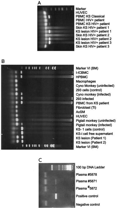
FIG. 4.
Detection of KSHV (HHV-8) DNA in various clinical samples from KS patients and infected cells. Genomic DNA was extracted with DNAzol reagent (Gibco-BRL, Gaithersburg, Md.), and 1 μg of total DNA was used in the PCR. For the detections of KSHV/HHV-8 expression, two primers were synthesized from the minor capsid region of the HHV-8 sequence. The sense primer was 5′-TCG AGC AGC TGT TGG TGT ACC ACA T, and the antisense primer was 5′-TCC GTG TTG TCT ACG TCC AG. These primers, used in the PCR, were designed to amplify 142 bp of HHV-8. Each PCR cycle consisted of denaturation at 94°C for 1 min, primer annealing at 60°C for 45 s, and extension at 72°C for 2 min. The samples were amplified for 30 cycles. A positive reaction for PCR of HHV-8 showed amplified product of 142 bp. Molecular weight markers (marker VI) were from Boehringer Mannheim, Indianapolis, Ind. (A) PBMCs from classic KS patients and HIV-infected KS patients, KS lesions, and skin from HIV-infected KS patients. (B) HHV-8-infected cells: PBMCs, monocytes, primary monkey peripheral blood cells, PBMCs from KS patients, the KS-1 cell line, which is persistently infected with HHV-8, and KS lesions from two patients. (C) HHV-8 DNA detected in plasma of two Israeli patients from Ethiopia.

