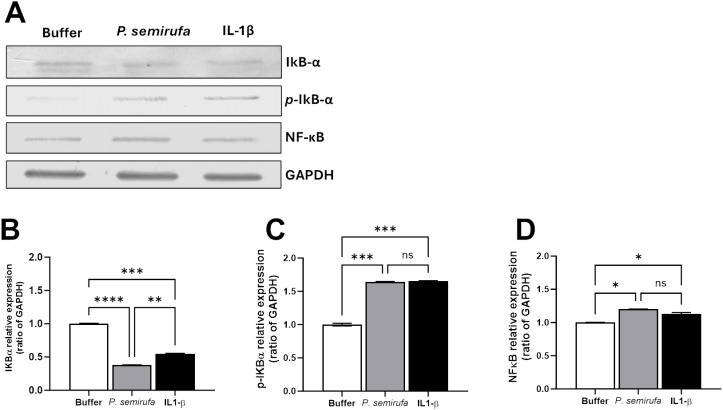
Fig 2. Western Blot and densitometric analysis of NF-κB, IκB-α, p-IκB-α, and GAPDH in chondrocytes.
Chondrocytes were treated with PBS buffer, IL-1β (10 ng/mL), or Pararama bristle extract (60 µg/mL). (A) Total protein extracts were prepared using RIPA lysis buffer containing protease inhibitors. Protein lysates (10 µg) were separated by 10% SDS-PAGE and transferred to nitrocellulose membranes. After blocking with PBS-BSA 5%, membranes were incubated with primary antibodies against IκB-α p-IκB-α, NF-κB, and GAPDH (all diluted 1:200). Following washes, membranes were treated with secondary antibodies (diluted 1:7500) and developed using NBT and BCIP (Promega, USA). GAPDH served as a loading control. (B-D) Densitometric analysis was conducted using ImageJ software to quantify the band intensity of target proteins relative to GAPDH. Data were analyzed using one-way ANOVA and Tukey’s post hoc test, with significance levels indicated as (*) p ≤ 0.05, (**) p ≤ 0.01, (***) p ≤ 0.001, and (****) p ≤ 0.0001.

