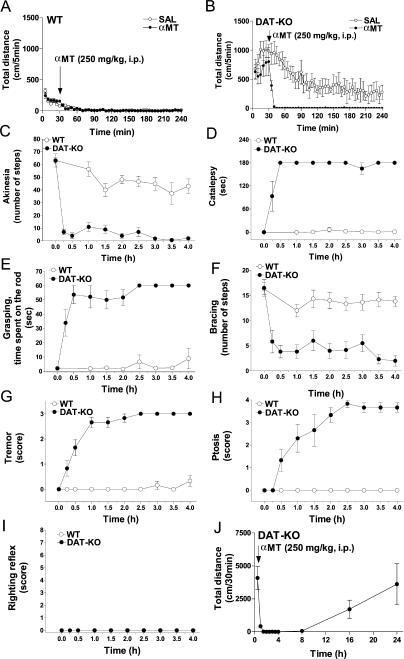
Figure 3. αMT-Induced Impairment in Motor Control in DAT-KO Mice.
Dynamics of locomotor activity following systemic administration of αMT (250 mg/kg IP) and saline (30 min after placement in the locomotor activity chamber) in WT (A) and DAT-KO (B) mice (n = 6–8 per group). Analysis of total distance traveled for 210 min after drug administration revealed significant effect of αMT treatment (p < 0.05; Student's t-test) in DAT-KO but not WT mice (WT-saline, 516 ± 50 cm/210 min; WT-αMT, 505 ± 98 cm/210 min; DAT-KO–saline, 18,489 ± 4,795 cm/210 min; DAT-KO–αMT, 448 ± 75 cm/210 min). αMT (injected at time 0) induced profound alterations in the akinesia (C), catalepsy (D), grasping (E), bracing (F) tremor (G), and ptosis (H) tests, but did not affect the righting reflex (I) in DAT-KO mice. Behavioral tests were performed as described in Materials and Methods. At all the time points, DAT-KO mice were significantly different versus respective values (data not shown) of saline-treated DAT-KO controls (p < 0.05; Student's t-test n = 6 per group) in these tests with exception of 15-min time point for ptosis (H) and all time points for righting reflex test (I). In WT mice only the akinesia test (C) revealed minor, yet significant, effect (1.5–4 h after αMT treatment) versus values (data not shown) of the respective saline treated WT controls (p < 0.05; Student's t-test; n = 6 per group). No significant alterations in any other test at any time point examined (D–I) was noted in αMT-treated versus saline treated (data not shown) WT mice. Locomotor activity is restored in DAT-KO mice 16–24h after αMT (250 mg/kg IP) treatment (J).

