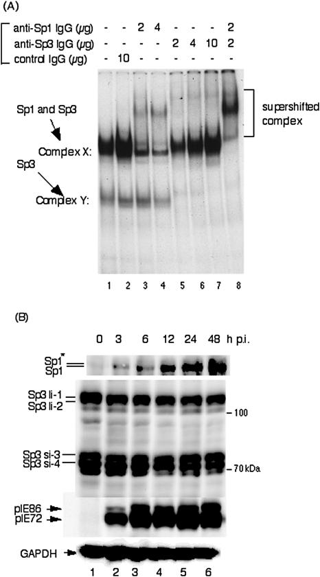
FIG. 1.
Sp1 and Sp3 transcription factors bind to GC boxes located in the proximal enhancer of HCMV MIE genes. (A) Supershift assay of DNA-protein complexes with anti-Sp1 and/or anti-Sp3 antibodies plus nuclear extracts using the 32P-labeled Sp1(−75) as a probe. Lane 1, probe plus nuclear extract; lane 2, probe plus nuclear extract with 10 μg of rabbit control IgG; lanes 3 and 4, probe plus nuclear extract with 2 and 4 μg of anti-Sp1 antibody, respectively; lanes 5, 6, and 7, probe plus nuclear extract with 2, 4, and 10 μg of anti-Sp3 antibody, respectively; lane 8, probe plus nuclear extract with 2 μg of anti-Sp1 and anti-Sp3 antibodies. Similar results were obtained using 32P-labeled Sp1(−55) as a probe.(B) Expression levels of Sp1 and Sp3 transcription factors after infection with HCMV. HFF cells were infected with HCMV at an MOI of 3 and harvested at the indicated times. Protein samples were prepared and 20 μg of each protein was applied for Western blot analyses. Proteins were separated by using 10% SDS-polyacrylamide gels. Detection of Sp1, Sp3, and immediate-early pIE72 (UL123)/pIE86 (UL122) was performed with polyclonal antibodies sc-59 and sc-644 and monoclonal antibody NEA-9221, respectively. Anti-pGAPDH antibody was used to confirm equal protein loading. For Sp1 the asterisk indicates the phosphorylated form. Abbreviations: Sp3li-1 and -2, long isoforms of Sp3; Sp3si-3 and -4, small isoforms of Sp3. The time p.i. is given at the top of each lane.

