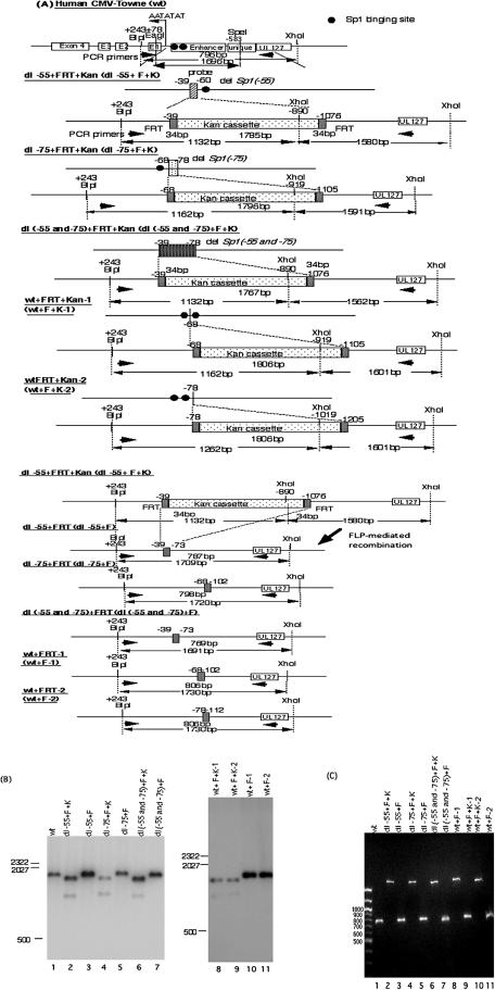
FIG. 4.
Structural analyses of recombinant HCMV BAC DNAs. (A) Schematic illustrations of parental and recombinant BAC DNAs with and without the Kanr gene. All recombinant BAC DNAs were constructed using a PCR-based rapid recombination system, and the Kanr gene was excised as described in Materials and Methods. The viral DNA fragment sizes of all recombinant viruses digested with restriction endonucleases BlpI and XhoI are indicated. Southern blot hybridization was performed with the 32P-labeled probe designated in the diagram for the human CMV Towne strain (wt). PCR was performed using primers designated in the diagram. (B) Southern blot analysis of the parental (wt) and dl−55+F, dl−75+F, dl(−55 and −75)+F, wt+F-1, and wt+F-2 recombinant BAC DNAs, with and without Kanr. Viral DNAs were digested with restriction endonucleases BlpI and XhoI, fractionated by electrophoresis in 1.0% agarose gels, and subjected to hybridization with the 32P-labeled probe. Standard molecular size markers are indicated in base pairs. BAC DNAs are identified at the tops of the blots. (C) PCR analysis of the parental and recombinant BAC DNAs. The products were amplified using the primer pair shown in panel A and fractionated by electrophoresis in 1.0% agarose and stained with ethidium bromide. BAC DNAs are identified at the top of the blot.

