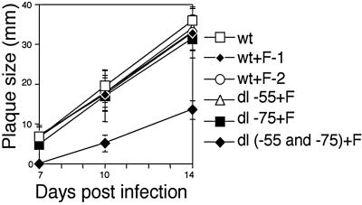
FIG. 7.
Plaque formation after infection with recombinant virus having both Sp1/Sp3 binding sites deleted. All plaques were generated from inocula infected with wt or wt+F-1, wt+F-2, dl-55+F, dl-75+F or dl(−55 and −75)+F recombinant viruses at an MOI of 0.01 in HFF cells. Each plaque size was determined as a mean of minimum and maximum lengths at the indicated days after infection. The results are averages for 10 plaques.

