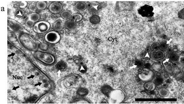
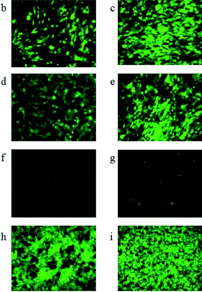
FIG. 4.
(a) Electron micrograph of CHO cells 24 h after infection with 5 PFU/cell HSV1716gfp/BHK. Examples of capsids in the nucleus (Nuc) are indicated by a black arrow, enveloped virions in the cytoplasm (Cyt) are indicated by a white arrow, and unenveloped capsids in the cytoplasm are indicated by a white arrowhead. Bar, 1 μm. For panels b to f, unprocessed images (magnification, ×200) from fluorescent microscopy are shown. (b) BHK cells after 8 h of infection with 5 PFU/cell HSV1716gfp propagated in CHO cells; (c) BHK cells after 8 h of infection with 5 PFU/cell HSV1716gfp propagated in Vero cells; (d) C8161 cells after 8 h of infection with 5 PFU/cell HSV1716gfp propagated in CHO cells; (e) BHK cells after 8 h of infection with 5 PFU/cell HSV1716gfp propagated in C8161 cells; (f) CHO cells after 8 h of infection with 5 PFU/cell HSV1716gfp propagated in CHO cells. For panels g, h, and i, unprocessed images (magnification, ×100) from fluorescent microscopy are shown for CHO (g), BHK (h), and Vero (i) cell lines 5 days after infection with 1 × 10³ PFU HSV1716gfp/BHK (g, h) or HSV1716gfp/CHO (i).


