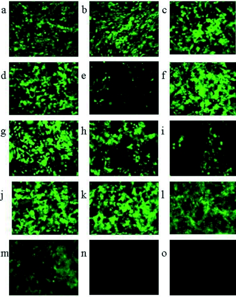
FIG. 5.
Unprocessed images (magnification, ×196) from fluorescent microscopy of various cell lines after 8 h of infection with HSV1716gfp or HSV17+gfp propagated either in BHK or Vero cells. For panels a to d, serum-starved or non-serum-starved BHK (a, b) or C8161 (c, d) cells were infected with 5 PFU/cell HSV1716gfp/BHK (a, b) or 5 PFU/cell HSV1716gfp/Vero (c, d) added either to non-serum-starved cells (b, d) or to cells simultaneously with the reintroduction of serum (a, c). For panels e to k, serum-starved or non-serum-starved CHO cells were infected with 5 PFU/cell HSV1716gfp/BHK added either to non-serum-starved cells (e) or to serum-starved cells simultaneously with the reintroduction of serum (f, j, k) or 4 (g), 8 (h), or 16 (i) hours after serum addition. For panels l and m, 5 PFU/cell HSV17+gfp/BHK was added to serum-starved CHO cells either simultaneously with serum addition (l) or 16 h after the reintroduction of serum (m). For panels n and o, 5 PFU/cell HSV1716gfp/Vero was added to either non-serum-starved CHO cells (n) or to serum-starved CHO cells simultaneously with the reintroduction of serum (o).

