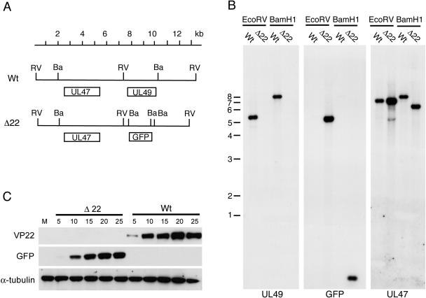
FIG. 2.
Characterization of the Δ22 virus. (A) DNA map of the HSV-1 genome across the UL49 gene, showing the relevant BamHI (Ba) and EcoRV (RV) sites. (B) Southern blotting of EcoRV- and BamHI-digested genomic DNA from WT and Δ22 viruses. The blots have been hybridized with probes for the UL49, GFP, and UL47 genes. (C) Monolayers of Vero cells were infected with either WT or Δ22 virus at a multiplicity of 10 and harvested at various time points ranging from 5 to 25 h. Equal amounts of total cell lysates were analyzed by SDS-PAGE followed by Western blotting with antibodies against VP22, GFP, and α-tubulin. Numbers to the left indicate DNA size markers in kilobases.

